

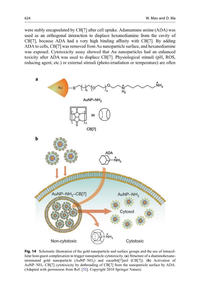
were stably encapsulated by CB[7] after cell uptake. Adamantane amine (ADA) was
used as an orthogonal interaction to displace hexanediamine from the cavity of
CB[7], because ADA had a very high binding affinity with CB[7]. By adding
ADA to cells, CB[7] was removed from Au nanoparticle surface, and hexanediamine
was exposed. Cytotoxicity assay showed that Au nanoparticles had an enhanced
toxicity after ADA was used to displace CB[7]. Physiological stimuli (pH, ROS,
reducing agent, etc.) or external stimuli (photo-irradiation or temperature) are often
Fig. 14 Schematic illustration of the gold nanoparticle and surface groups and the use of intracellular host-guest complexation to trigger nanoparticle cytotoxicity. (a) Structure of a diaminohexaneterminated gold nanoparticle (AuNP–NH 2 ) and cucurbit[7]uril (CB[7]). (b) Activation of
AuNP–NH 2 –CB[7] cytotoxicity by dethreading of CB[7] from the nanoparticle surface by ADA.
(Adapted with permission from Ref. [32]. Copyright 2010 Springer Nature)
624
W. Mao and D. Ma