

78
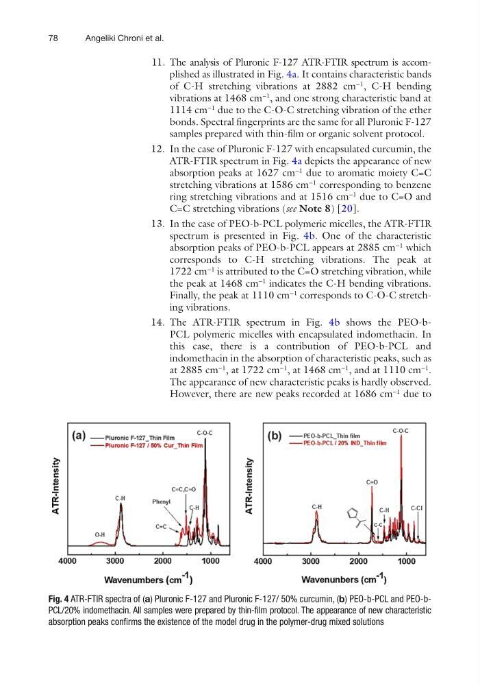
11. The analysis of Pluronic F-127 ATR-FTIR spectrum is accomplished as illustrated in Fig. 4a. It contains characteristic bands
of C-H stretching vibrations at 2882 cm
−1
, C-H bending
vibrations at 1468 cm
−1
, and one strong characteristic band at
1114 cm
−1
due to the C-O-C stretching vibration of the ether
bonds. Spectral fingerprints are the same for all Pluronic F-127
samples prepared with thin-film or organic solvent protocol.
12. In the case of Pluronic F-127 with encapsulated curcumin, the
ATR-FTIR spectrum in Fig. 4a depicts the appearance of new
absorption peaks at 1627 cm
−1
due to aromatic moiety C=C
stretching vibrations at 1586 cm
−1
corresponding to benzene
ring stretching vibrations and at 1516 cm
−1
due to C=O and
C=C stretching vibrations (see Note 8) [20].
13. In the case of PEO-b-PCL polymeric micelles, the ATR-FTIR
spectrum is presented in Fig. 4b. One of the characteristic
absorption peaks of PEO-b-PCL appears at 2885 cm
−1
which
corresponds to C-H stretching vibrations. The peak at
1722 cm
−1
is attributed to the C=O stretching vibration, while
the peak at 1468 cm
−1
indicates the C-H bending vibrations.
Finally, the peak at 1110 cm
−1
corresponds to C-O-C stretching vibrations.
14. The ATR-FTIR spectrum in Fig. 4b shows the PEO-bPCL polymeric micelles with encapsulated indomethacin. In
this case, there is a contribution of PEO-b-PCL and
indomethacin in the absorption of characteristic peaks, such as
at 2885 cm
−1
, at 1722 cm
−1
, at 1468 cm
−1
, and at 1110 cm
−1
.
The appearance of new characteristic peaks is hardly observed.
However, there are new peaks recorded at 1686 cm
−1
due to
Fig. 4 ATR-FTIR spectra of (a) Pluronic F-127 and Pluronic F-127/ 50% curcumin, (b) PEO-b-PCL and PEO-bPCL/20% indomethacin. All samples were prepared by thin-film protocol. The appearance of new characteristic
absorption peaks confirms the existence of the model drug in the polymer-drug mixed solutions
Angeliki Chroni et al.
11. The analysis of Pluronic F-127 ATR-FTIR spectrum is accomplished as illustrated in Fig. 4a. It contains characteristic bands
of C-H stretching vibrations at 2882 cm
−1
, C-H bending
vibrations at 1468 cm
−1
, and one strong characteristic band at
1114 cm
−1
due to the C-O-C stretching vibration of the ether
bonds. Spectral fingerprints are the same for all Pluronic F-127
samples prepared with thin-film or organic solvent protocol.
12. In the case of Pluronic F-127 with encapsulated curcumin, the
ATR-FTIR spectrum in Fig. 4a depicts the appearance of new
absorption peaks at 1627 cm
−1
due to aromatic moiety C=C
stretching vibrations at 1586 cm
−1
corresponding to benzene
ring stretching vibrations and at 1516 cm
−1
due to C=O and
C=C stretching vibrations (see Note 8) [20].
13. In the case of PEO-b-PCL polymeric micelles, the ATR-FTIR
spectrum is presented in Fig. 4b. One of the characteristic
absorption peaks of PEO-b-PCL appears at 2885 cm
−1
which
corresponds to C-H stretching vibrations. The peak at
1722 cm
−1
is attributed to the C=O stretching vibration, while
the peak at 1468 cm
−1
indicates the C-H bending vibrations.
Finally, the peak at 1110 cm
−1
corresponds to C-O-C stretching vibrations.
14. The ATR-FTIR spectrum in Fig. 4b shows the PEO-bPCL polymeric micelles with encapsulated indomethacin. In
this case, there is a contribution of PEO-b-PCL and
indomethacin in the absorption of characteristic peaks, such as
at 2885 cm
−1
, at 1722 cm
−1
, at 1468 cm
−1
, and at 1110 cm
−1
.
The appearance of new characteristic peaks is hardly observed.
However, there are new peaks recorded at 1686 cm
−1
due to
Fig. 4 ATR-FTIR spectra of (a) Pluronic F-127 and Pluronic F-127/ 50% curcumin, (b) PEO-b-PCL and PEO-bPCL/20% indomethacin. All samples were prepared by thin-film protocol. The appearance of new characteristic
absorption peaks confirms the existence of the model drug in the polymer-drug mixed solutions
Angeliki Chroni et al.