

77
1. Select the preferable measurement options, e.g., spectral range
from 5000 to 500 cm
−1
, wavenumber resolution of 4 cm
−1
and
number of scans 64.
2. Firstly, take a background before sample measurement.
3. Using a micropipette, add 20 μL of polymer solution directly
on the surface of the flat diamond crystal.
4. Using nitrogen gas flow, the polymer solution is purged at
100 ml/min flow rate to evaporate the solvent and the recording of the spectrum starts simultaneously.
5. FTIR-ATR spectra are recorded in real-time mode until
solvent evaporation occurs and a polymeric thin film is formed
on the diamond’s flat surface.
6. Remove the formed thin film from the surface of the flat diamond crystal by cleaning carefully the surface with pure H 2 O.
7. Perform again a new measurement without the deposition of
sample to clarify that the surface is clean as the obtained ATRFTIR spectrum indicates.
8. For the measurements of polymeric micelles with the encapsulated drug, add 20 μL solution directly on the surface of the
flat diamond crystal.
9. Follow steps 4–8 and repeat the procedure with the same
measurement conditions.
10. The spectra obtained by ATR-FTIR spectroscopy certify the
expected chemical structure of the block copolymers and the
successful encapsulation of the drug into polymeric micelles
(see Note 7).
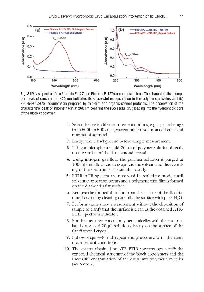
Fig. 3 UV-Vis spectra of (a) Pluronic F-127 and Pluronic F-127/curcumin solutions. The characteristic absorption peak of curcumin at 420 nm indicates its successful encapsulation in the polymeric micelles and (b)
PEO- b-PCL/20% indomethacin prepared by thin-film and organic solvent protocols. The observation of the
characteristic peak of indomethacin at 260 nm confirms the successful drug loading into the hydrophobic core
of the block copolymer
Drug Delivery: Hydrophobic Drug Encapsulation into Amphiphilic Block…
1. Select the preferable measurement options, e.g., spectral range
from 5000 to 500 cm
−1
, wavenumber resolution of 4 cm
−1
and
number of scans 64.
2. Firstly, take a background before sample measurement.
3. Using a micropipette, add 20 μL of polymer solution directly
on the surface of the flat diamond crystal.
4. Using nitrogen gas flow, the polymer solution is purged at
100 ml/min flow rate to evaporate the solvent and the recording of the spectrum starts simultaneously.
5. FTIR-ATR spectra are recorded in real-time mode until
solvent evaporation occurs and a polymeric thin film is formed
on the diamond’s flat surface.
6. Remove the formed thin film from the surface of the flat diamond crystal by cleaning carefully the surface with pure H 2 O.
7. Perform again a new measurement without the deposition of
sample to clarify that the surface is clean as the obtained ATRFTIR spectrum indicates.
8. For the measurements of polymeric micelles with the encapsulated drug, add 20 μL solution directly on the surface of the
flat diamond crystal.
9. Follow steps 4–8 and repeat the procedure with the same
measurement conditions.
10. The spectra obtained by ATR-FTIR spectroscopy certify the
expected chemical structure of the block copolymers and the
successful encapsulation of the drug into polymeric micelles
(see Note 7).
Fig. 3 UV-Vis spectra of (a) Pluronic F-127 and Pluronic F-127/curcumin solutions. The characteristic absorption peak of curcumin at 420 nm indicates its successful encapsulation in the polymeric micelles and (b)
PEO- b-PCL/20% indomethacin prepared by thin-film and organic solvent protocols. The observation of the
characteristic peak of indomethacin at 260 nm confirms the successful drug loading into the hydrophobic core
of the block copolymer
Drug Delivery: Hydrophobic Drug Encapsulation into Amphiphilic Block…