

309
Contrariwise, in negatively charged DMPC:DPPG membranes (Fig. 5) there was an increase of ΔT 1/2 , implying a
reduction of the cooperativity of the materials due to the
reduction of the membrane rigidity, which was more
intense in the presence of CPD G3 rather than CPD G4.
Although a reduction of cooperativity had taken place, no
phase separation was observed even at high CPD concentration. The observed concentration- dependent increase
of the fluidity of the DMPC:DPPG bilayer can be correlated with the negative charge of the bilayer, due to stronger dendrimer–lipid interaction via electrostatic forces.
10
12 14 16 18
20 22 24 26 28 30 32
10
12 14 16 18
20 22 24 26 28 30 32
a
b
c
d
e
a
b
c
d
e
Heat flow, Endotherm Heat flow, Endotherm
I
II
V h = 2
o C/min
ºC
ºC
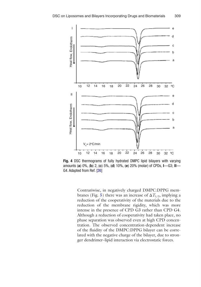
Fig. 4 DSC thermograms of fully hydrated DMPC lipid bilayers with varying
amounts (a) 0%, (b) 2, (c) 5%, (d) 10%, (e) 20% (molar) of CPDs, I—G3; II—
G4. Adapted from Ref. [26]
DSC on Liposomes and Bilayers Incorporating Drugs and Biomaterials
Contrariwise, in negatively charged DMPC:DPPG membranes (Fig. 5) there was an increase of ΔT 1/2 , implying a
reduction of the cooperativity of the materials due to the
reduction of the membrane rigidity, which was more
intense in the presence of CPD G3 rather than CPD G4.
Although a reduction of cooperativity had taken place, no
phase separation was observed even at high CPD concentration. The observed concentration- dependent increase
of the fluidity of the DMPC:DPPG bilayer can be correlated with the negative charge of the bilayer, due to stronger dendrimer–lipid interaction via electrostatic forces.
10
12 14 16 18
20 22 24 26 28 30 32
10
12 14 16 18
20 22 24 26 28 30 32
a
b
c
d
e
a
b
c
d
e
Heat flow, Endotherm Heat flow, Endotherm
I
II
V h = 2
o C/min
ºC
ºC
Fig. 4 DSC thermograms of fully hydrated DMPC lipid bilayers with varying
amounts (a) 0%, (b) 2, (c) 5%, (d) 10%, (e) 20% (molar) of CPDs, I—G3; II—
G4. Adapted from Ref. [26]
DSC on Liposomes and Bilayers Incorporating Drugs and Biomaterials