

180
1. Perform the characterization of compounds via
1
H-NMR and
13
C-NMR spectroscopy.
2. Dissolve 2 mg of each compound in 500 uL D 2 O in an
Eppendorf.
3. Transfer samples to 5 mm NMR tubes.
4. Load the sample into the Bruker AV-400 spectrometer.
5. Lock, tune, and match the sample according to the manufacturer’s guidelines (Fig. 2).
1. Dilute TMZ in a proper volume of MeOH to obtain the desired
concentration (seeNote 2).
2. Prepare aliquots of stock solutions from the initial stock of
TMZ (5–30 μM).
3. Scan samples by UV-Vis spectrophotometer in the range of
200–450 nm, using MeOH as blank.
4. Analyze samples for their respective absorbance at 330 nm λ max .
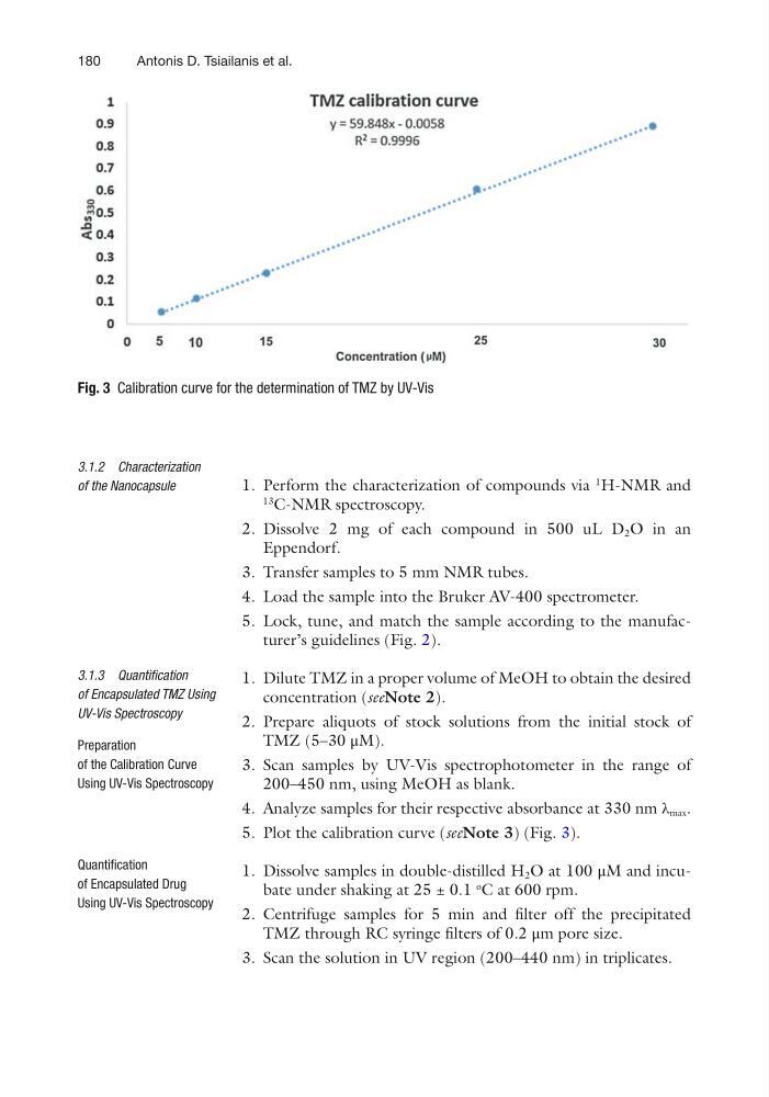
5. Plot the calibration curve (seeNote 3) (Fig. 3).
1. Dissolve samples in double-distilled H 2 O at 100 μM and incubate under shaking at 25 ± 0.1
ο
C at 600 rpm.
2. Centrifuge samples for 5 min and filter off the precipitated
TMZ through RC syringe filters of 0.2 μm pore size.
3. Scan the solution in UV region (200–440 nm) in triplicates.
3.1.2 Characterization
of the Nanocapsule
3.1.3 Quantification
of Encapsulated TMZ Using
UV-Vis Spectroscopy
Preparation
of the Calibration Curve
Using UV-Vis Spectroscopy
Quantification
of Encapsulated Drug
Using UV-Vis Spectroscopy
Fig. 3 Calibration curve for the determination of TMZ by UV-Vis
Antonis D. Tsiailanis et al.
1. Perform the characterization of compounds via
1
H-NMR and
13
C-NMR spectroscopy.
2. Dissolve 2 mg of each compound in 500 uL D 2 O in an
Eppendorf.
3. Transfer samples to 5 mm NMR tubes.
4. Load the sample into the Bruker AV-400 spectrometer.
5. Lock, tune, and match the sample according to the manufacturer’s guidelines (Fig. 2).
1. Dilute TMZ in a proper volume of MeOH to obtain the desired
concentration (seeNote 2).
2. Prepare aliquots of stock solutions from the initial stock of
TMZ (5–30 μM).
3. Scan samples by UV-Vis spectrophotometer in the range of
200–450 nm, using MeOH as blank.
4. Analyze samples for their respective absorbance at 330 nm λ max .
5. Plot the calibration curve (seeNote 3) (Fig. 3).
1. Dissolve samples in double-distilled H 2 O at 100 μM and incubate under shaking at 25 ± 0.1
ο
C at 600 rpm.
2. Centrifuge samples for 5 min and filter off the precipitated
TMZ through RC syringe filters of 0.2 μm pore size.
3. Scan the solution in UV region (200–440 nm) in triplicates.
3.1.2 Characterization
of the Nanocapsule
3.1.3 Quantification
of Encapsulated TMZ Using
UV-Vis Spectroscopy
Preparation
of the Calibration Curve
Using UV-Vis Spectroscopy
Quantification
of Encapsulated Drug
Using UV-Vis Spectroscopy
Fig. 3 Calibration curve for the determination of TMZ by UV-Vis
Antonis D. Tsiailanis et al.