

158
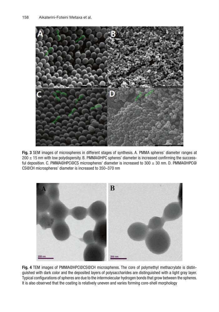
Fig. 3 SEM images of microspheres in different stages of synthesis. A. PMMA spheres’ diameter ranges at
200 ± 15 nm with low polydispersity. B. PMMA@HPC spheres’ diameter is increased confirming the successful deposition. C. PMMA@HPC@CS microspheres’ diameter is increased to 300 ± 30 nm. D. PMMA@HPC@
CS@CH microspheres’ diameter is increased to 350–370 nm
Fig. 4 TEM images of PMMA@HPC@CS@CH microspheres. The core of polymethyl methacrylate is distinguished with dark color and the deposited layers of polysaccharides are distinguished with a light gray layer.
Typical configurations of spheres are due to the intermolecular hydrogen bonds that grow between the spheres.
It is also observed that the coating is relatively uneven and varies forming core-shell morphology
Aikaterini-Foteini Metaxa et al.
Fig. 3 SEM images of microspheres in different stages of synthesis. A. PMMA spheres’ diameter ranges at
200 ± 15 nm with low polydispersity. B. PMMA@HPC spheres’ diameter is increased confirming the successful deposition. C. PMMA@HPC@CS microspheres’ diameter is increased to 300 ± 30 nm. D. PMMA@HPC@
CS@CH microspheres’ diameter is increased to 350–370 nm
Fig. 4 TEM images of PMMA@HPC@CS@CH microspheres. The core of polymethyl methacrylate is distinguished with dark color and the deposited layers of polysaccharides are distinguished with a light gray layer.
Typical configurations of spheres are due to the intermolecular hydrogen bonds that grow between the spheres.
It is also observed that the coating is relatively uneven and varies forming core-shell morphology
Aikaterini-Foteini Metaxa et al.