

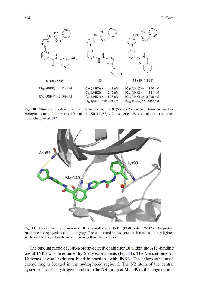
The binding mode of JNK-isoform-selective inhibitor 10 within the ATP-binding
site of JNK3 was determined by X-ray experiments (Fig. 11). The R-enantiomer of
10 forms several hydrogen bond interactions with JNK3. The chloro-substituted
phenyl ring is located in the hydrophobic region I. The N2 atom of the central
pyrazole accepts a hydrogen bond from the NH group of Met149 of the hinge region.
Fig. 10 Structural modifications of the lead structure 9 (SR-4326) and structures as well as
biological data of inhibitors 10 and 11 (SR-11935) of this series. Biological data are taken
from Zheng et al. [37]
Fig. 11 X-ray structure of inhibitor 10 in complex with JNK3 (PDB code: 4WHZ). The protein
backbone is displayed as cartoon in gray. The compound and selected amino acids are highlighted
as sticks. Hydrogen bonds are shown as yellow dashed lines
214
P. Koch
site of JNK3 was determined by X-ray experiments (Fig. 11). The R-enantiomer of
10 forms several hydrogen bond interactions with JNK3. The chloro-substituted
phenyl ring is located in the hydrophobic region I. The N2 atom of the central
pyrazole accepts a hydrogen bond from the NH group of Met149 of the hinge region.
Fig. 10 Structural modifications of the lead structure 9 (SR-4326) and structures as well as
biological data of inhibitors 10 and 11 (SR-11935) of this series. Biological data are taken
from Zheng et al. [37]
Fig. 11 X-ray structure of inhibitor 10 in complex with JNK3 (PDB code: 4WHZ). The protein
backbone is displayed as cartoon in gray. The compound and selected amino acids are highlighted
as sticks. Hydrogen bonds are shown as yellow dashed lines
214
P. Koch