

therefore considered highly unstable [83]. Interestingly, this water molecule (that we
will call Water1) was computed to be located in proximity to the 2-position of the
indoline core and equi-energetically present in PI3Kδ (+5.8 kcal/mol). Visual
inspection of the 3D structure established by WaterMap suggested that any substituent in the indoline 2-position with R or “down” stereochemistry would be perfectly
suited for displacing Water1 leading to gain in free energy of ligand binding.
WaterMap calculations with (S)-27 recapitulated the same observation as for 24;
an unstable water molecule (Water1) is trapped by the (S)-ligand in the active site
(Fig. 19b) decreasing its affinity (ΔG) vs the (R)-enantiomer which displaces it
(Table 3). To reinforce the validity of this hypothesis, it is worth mentioning that
computed free energies of binding were aligned in trends with the experimental ITC
values collected (data not shown) not only for 24 and 27 but for a series of
2-substituted indoline analogs [83].
When applied to PI3Kδ, Water1 was not only retrieved but also estimated to be
even more instable (+6.7 kcal/mol vs +5.9 kcal/mol in the case of (S)-27). However,
the respective free energy values are too similar to quantitatively explain the
observed selectivity pattern of the two enantiomers. A favorable displacement of
the water molecule in PI3Kδ compared to PI3kβ cannot solely explain why (R)-27 is
more potent and less selective.
To better assess the origin of this selectivity pattern, binding site water molecules
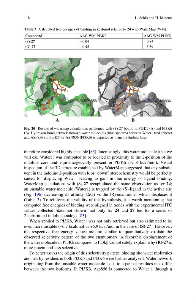
and nearby residues in both PI3Kβ and PI3Kδ were further analyzed. Water network
originating from the unstable water molecule leads to a pair of residues that differ
between the two isoforms. In PI3Kβ, Asp856 is connected to Water 1 through a
Table 3 Calculated free energies of binding in kcal/mol relative to 24 with WaterMap (WM)
Compound
ΔΔG WM PI3Kβ
Δ Δ G WM PI3Kδ
(S)-27
À0.04
0.84
(R)-27
À0.45
À3.59
Fig. 20 Results of watermap calculations performed with (S)-27 bound to PI3Kβ (A) and PI3Kδ
(B). Hydrogen-bond network through water molecules (blue spheres) between Water1 (red sphere)
and ASP856 (in PI3Kβ) or ASN836 (PI3Kδ) is depicted as magenta dashed lines
118
L. Schio and H. Minoux
will call Water1) was computed to be located in proximity to the 2-position of the
indoline core and equi-energetically present in PI3Kδ (+5.8 kcal/mol). Visual
inspection of the 3D structure established by WaterMap suggested that any substituent in the indoline 2-position with R or “down” stereochemistry would be perfectly
suited for displacing Water1 leading to gain in free energy of ligand binding.
WaterMap calculations with (S)-27 recapitulated the same observation as for 24;
an unstable water molecule (Water1) is trapped by the (S)-ligand in the active site
(Fig. 19b) decreasing its affinity (ΔG) vs the (R)-enantiomer which displaces it
(Table 3). To reinforce the validity of this hypothesis, it is worth mentioning that
computed free energies of binding were aligned in trends with the experimental ITC
values collected (data not shown) not only for 24 and 27 but for a series of
2-substituted indoline analogs [83].
When applied to PI3Kδ, Water1 was not only retrieved but also estimated to be
even more instable (+6.7 kcal/mol vs +5.9 kcal/mol in the case of (S)-27). However,
the respective free energy values are too similar to quantitatively explain the
observed selectivity pattern of the two enantiomers. A favorable displacement of
the water molecule in PI3Kδ compared to PI3kβ cannot solely explain why (R)-27 is
more potent and less selective.
To better assess the origin of this selectivity pattern, binding site water molecules
and nearby residues in both PI3Kβ and PI3Kδ were further analyzed. Water network
originating from the unstable water molecule leads to a pair of residues that differ
between the two isoforms. In PI3Kβ, Asp856 is connected to Water 1 through a
Table 3 Calculated free energies of binding in kcal/mol relative to 24 with WaterMap (WM)
Compound
ΔΔG WM PI3Kβ
Δ Δ G WM PI3Kδ
(S)-27
À0.04
0.84
(R)-27
À0.45
À3.59
Fig. 20 Results of watermap calculations performed with (S)-27 bound to PI3Kβ (A) and PI3Kδ
(B). Hydrogen-bond network through water molecules (blue spheres) between Water1 (red sphere)
and ASP856 (in PI3Kβ) or ASN836 (PI3Kδ) is depicted as magenta dashed lines
118
L. Schio and H. Minoux