

patients which triggered the decision to evaluate the drug in a phase II clinical trial in
MET-amplified NSCLC patients (NCT02435121) [56].
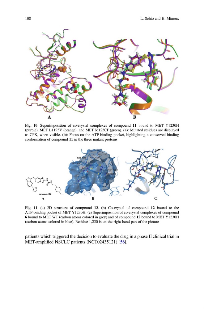
Fig. 10 Superimposition of co-crystal complexes of compound 11 bound to MET Y1230H
(purple), MET L1195V (orange), and MET M1250T (green). (a): Mutated residues are displayed
as CPK, when visible. (b): Focus on the ATP-binding pocket, highlighting a conserved binding
conformation of compound 11 in the three mutant proteins
S
N
S
N
N
N
N
S
N
H
N
H
O
N
compound 12
A
B
C
Fig. 11 (a) 2D structure of compound 12. (b) Co-crystal of compound 12 bound to the
ATP-binding pocket of MET Y1230H. (c) Superimposition of co-crystal complexes of compound
6 bound to MET WT (carbon atoms colored in grey) and of compound 12 bound to MET Y1230H
(carbon atoms colored in blue). Residue 1,230 is on the right-hand part of the picture
108
L. Schio and H. Minoux
MET-amplified NSCLC patients (NCT02435121) [56].
Fig. 10 Superimposition of co-crystal complexes of compound 11 bound to MET Y1230H
(purple), MET L1195V (orange), and MET M1250T (green). (a): Mutated residues are displayed
as CPK, when visible. (b): Focus on the ATP-binding pocket, highlighting a conserved binding
conformation of compound 11 in the three mutant proteins
S
N
S
N
N
N
N
S
N
H
N
H
O
N
compound 12
A
B
C
Fig. 11 (a) 2D structure of compound 12. (b) Co-crystal of compound 12 bound to the
ATP-binding pocket of MET Y1230H. (c) Superimposition of co-crystal complexes of compound
6 bound to MET WT (carbon atoms colored in grey) and of compound 12 bound to MET Y1230H
(carbon atoms colored in blue). Residue 1,230 is on the right-hand part of the picture
108
L. Schio and H. Minoux