

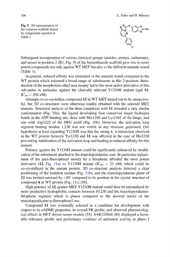
Subsequent incorporation of various chemical groups (amides, amines, carbamates,
and ureas) in position 2 (R1, Fig. 9) of the benzothiazole scaffold gave rise to more
potent compounds not only against WT MET but also vs the different mutants tested
(Table 1).
In general, reduced affinity was measured vs the mutants tested compared to the
WT protein which tolerated a broad range of substituents in this 2-position. Introduction of the morpholino-ethyl urea moiety led to the most active derivative of this
sub-series in particular against the clinically relevant Y1230H mutant (cpd 11;
IC 50 ¼ 204 nM).
Attempts to co-crystallize compound 11 in WT MET turned out to be unsuccessful, but 3D co-structures were otherwise readily obtained with the selected MET
mutants. Structural analysis of the three complexes with 11 revealed a very similar
conformation (Fig. 10a), the ligand developing four conserved major hydrogen
bonds in the ATP binding site, three with Met1160 and Lys1161 of the hinge, and
one with Asp1222 of the DFG motif (Fig. 10b). However, the activation loop
segment bearing residue 1,230 was not visible in any structure generated. Our
hypothesis at least regarding Y1230H was that the strong π- π interaction observed
in the WT protein between Tyr1230 and 11 was affected in the case of His1230
preventing stabilization of the activation loop and leading to reduced affinity for this
mutant.
Potency against the Y1230H mutant could be significantly enhanced by modification of the substituent attached to the triazolopyridazine core. In particular replacement of the para-fluorophenyl moiety by a thiophene afforded the most potent
derivative (12, Fig. 11a) vs Y1230H mutant (IC 50 ¼ 23 nM) which could be
co-crystallized in the mutant protein. 3D co-structure analysis detected a clear
positioning of the histidine residue (Fig. 11b), and the triazolopyridazine plane of
12 was twisted outward by ~20
compared to its position in the crystal structure of
compound 6 in WT protein (Fig. 11c) [50].
High potency of 12 against MET Y1230H mutant could then be rationalized by
more productive hydrophobic contacts between H1230 and the triazolopyridazinethiophene segment which is planar compared to the skewed nature of the
triazolopyridazine-p-fluorophenyl one.
Compound 11 was eventually selected as a candidate for development with
respect to its eADME properties, its overall PK profile, and observed pharmacological effects in MET driven tumor models [55]. SAR125844 (11) displayed a favorable tolerance profile and preliminary evidence of antitumor activity in phase I
N
N
N
N
S
F
N
S
R1
Fig. 9 2D representation of
the common scaffold shared
by compounds reported in
Table 1
106
L. Schio and H. Minoux
and ureas) in position 2 (R1, Fig. 9) of the benzothiazole scaffold gave rise to more
potent compounds not only against WT MET but also vs the different mutants tested
(Table 1).
In general, reduced affinity was measured vs the mutants tested compared to the
WT protein which tolerated a broad range of substituents in this 2-position. Introduction of the morpholino-ethyl urea moiety led to the most active derivative of this
sub-series in particular against the clinically relevant Y1230H mutant (cpd 11;
IC 50 ¼ 204 nM).
Attempts to co-crystallize compound 11 in WT MET turned out to be unsuccessful, but 3D co-structures were otherwise readily obtained with the selected MET
mutants. Structural analysis of the three complexes with 11 revealed a very similar
conformation (Fig. 10a), the ligand developing four conserved major hydrogen
bonds in the ATP binding site, three with Met1160 and Lys1161 of the hinge, and
one with Asp1222 of the DFG motif (Fig. 10b). However, the activation loop
segment bearing residue 1,230 was not visible in any structure generated. Our
hypothesis at least regarding Y1230H was that the strong π- π interaction observed
in the WT protein between Tyr1230 and 11 was affected in the case of His1230
preventing stabilization of the activation loop and leading to reduced affinity for this
mutant.
Potency against the Y1230H mutant could be significantly enhanced by modification of the substituent attached to the triazolopyridazine core. In particular replacement of the para-fluorophenyl moiety by a thiophene afforded the most potent
derivative (12, Fig. 11a) vs Y1230H mutant (IC 50 ¼ 23 nM) which could be
co-crystallized in the mutant protein. 3D co-structure analysis detected a clear
positioning of the histidine residue (Fig. 11b), and the triazolopyridazine plane of
12 was twisted outward by ~20
compared to its position in the crystal structure of
compound 6 in WT protein (Fig. 11c) [50].
High potency of 12 against MET Y1230H mutant could then be rationalized by
more productive hydrophobic contacts between H1230 and the triazolopyridazinethiophene segment which is planar compared to the skewed nature of the
triazolopyridazine-p-fluorophenyl one.
Compound 11 was eventually selected as a candidate for development with
respect to its eADME properties, its overall PK profile, and observed pharmacological effects in MET driven tumor models [55]. SAR125844 (11) displayed a favorable tolerance profile and preliminary evidence of antitumor activity in phase I
N
N
N
N
S
F
N
S
R1
Fig. 9 2D representation of
the common scaffold shared
by compounds reported in
Table 1
106
L. Schio and H. Minoux