

triazolopyridazine core, a chemical group introduced in this benzimidazole series in
a historical anti-helminthic project developed in the 1980s in the company. Compound 6 could be co-crystallized in MET, and the 3D-structure of the complex was
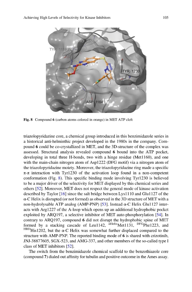
assessed. Structural analysis revealed compound 6 bound into the ATP pocket,
developing in total three H-bonds, two with a hinge residue (Met1160), and one
with the main-chain nitrogen atom of Asp1222 (DFG motif) via a nitrogen atom of
the triazolopyridazine moiety. Moreover, the triazolopyridazine ring made a specific
π-π interaction with Tyr1230 of the activation loop found in a non-competent
conformation (Fig. 8). This specific binding mode involving Tyr1230 is believed
to be a major driver of the selectivity for MET displayed by this chemical series and
others [52]. Moreover, MET does not respect the general mode of kinase activation
described by Taylor [16] since the salt bridge between Lys1110 and Glu1127 of the
α-C Helix is disrupted (or not formed) as observed in the 3D structure of MET with a
non-hydrolysable ATP analog (AMP-PNP) [53]. Instead α-C Helix Glu1127 interacts with Arg1227 of the A-loop which opens up an additional hydrophobic pocket
exploited by ARQ197, a selective inhibitor of MET auto-phosphorylation [54]. In
contrary to ARQ197, compound 6 did not disrupt the hydrophobic spine of MET
formed by a stacking cascade of Leu1142,
αHelix Met1131,
DFG
Phe1223, and
HRD His1202, but the α-C Helix was somewhat further displaced compared to the
structure with AMP-PNP. The reported binding mode of 6 is shared with crizotinib,
JNJ-38877605, SGX-523, and AMG-337, and other members of the so-called type I
class of MET inhibitors [52].
The switch from the benzimidazole chemical scaffold to the benzothiazole core
(compound 7) dialed out affinity for tubulin and positive outcome in the Ames assay.
Fig. 8 Compound 6 (carbon atoms colored in orange) in MET ATP cleft
Achieving High Levels of Selectivity for Kinase Inhibitors
105
a historical anti-helminthic project developed in the 1980s in the company. Compound 6 could be co-crystallized in MET, and the 3D-structure of the complex was
assessed. Structural analysis revealed compound 6 bound into the ATP pocket,
developing in total three H-bonds, two with a hinge residue (Met1160), and one
with the main-chain nitrogen atom of Asp1222 (DFG motif) via a nitrogen atom of
the triazolopyridazine moiety. Moreover, the triazolopyridazine ring made a specific
π-π interaction with Tyr1230 of the activation loop found in a non-competent
conformation (Fig. 8). This specific binding mode involving Tyr1230 is believed
to be a major driver of the selectivity for MET displayed by this chemical series and
others [52]. Moreover, MET does not respect the general mode of kinase activation
described by Taylor [16] since the salt bridge between Lys1110 and Glu1127 of the
α-C Helix is disrupted (or not formed) as observed in the 3D structure of MET with a
non-hydrolysable ATP analog (AMP-PNP) [53]. Instead α-C Helix Glu1127 interacts with Arg1227 of the A-loop which opens up an additional hydrophobic pocket
exploited by ARQ197, a selective inhibitor of MET auto-phosphorylation [54]. In
contrary to ARQ197, compound 6 did not disrupt the hydrophobic spine of MET
formed by a stacking cascade of Leu1142,
αHelix Met1131,
DFG
Phe1223, and
HRD His1202, but the α-C Helix was somewhat further displaced compared to the
structure with AMP-PNP. The reported binding mode of 6 is shared with crizotinib,
JNJ-38877605, SGX-523, and AMG-337, and other members of the so-called type I
class of MET inhibitors [52].
The switch from the benzimidazole chemical scaffold to the benzothiazole core
(compound 7) dialed out affinity for tubulin and positive outcome in the Ames assay.
Fig. 8 Compound 6 (carbon atoms colored in orange) in MET ATP cleft
Achieving High Levels of Selectivity for Kinase Inhibitors
105