

contact of the αC-helix to the ATP cleft. This conformation called αC-helix out has
been also reported to be the very specific inhibitory mechanism of lapatinib against
Her2 and EGFR [30].
Despite decent potency vs Aurora A, compound 3 was devoid of any antiproliferative activity when tested against a limited panel of cancer cell lines. Subsequent gain of affinity was obtained by removing the nitrogen atom in the pyridinone
ring despite structural evidence of its involvement in an H-bond with a water
molecule in the binding pocket. The resulting molecule (compound 4, Fig. 5), a
low double digit nanomolar Aurora A inhibitor, blocked Hela proliferation at
300 nM (IC 50 ). Compound 4 exhibited also a potent affinity for Aurora B
(IC 50 ¼ 5 nM) and otherwise was shown to be totally inactive against other kinases
tested (except Tie2, IC 50 ¼ 3 μM).
In a second round of chemical optimization, the pyrazole moiety was modified,
and the 2-ethoxycarbonyl-pyrrole group emerged as being the most productive
substitution in terms of anti-proliferative potency in Hela cells (compound 5,
Fig. 5), whereas biochemical activity was only slightly improved compared to 4.
Compound 5 was resolved by chiral chromatography, and the dextrogyre enantiomer
(+)-5 (S-configuration) was identified as the active substance of the racemic mixture:
Aurora A, IC 50 ¼ 4 nM; Aurora B, IC 50 ¼ 2 nM; and Hela cells, IC 50 ¼ 2 nM (the
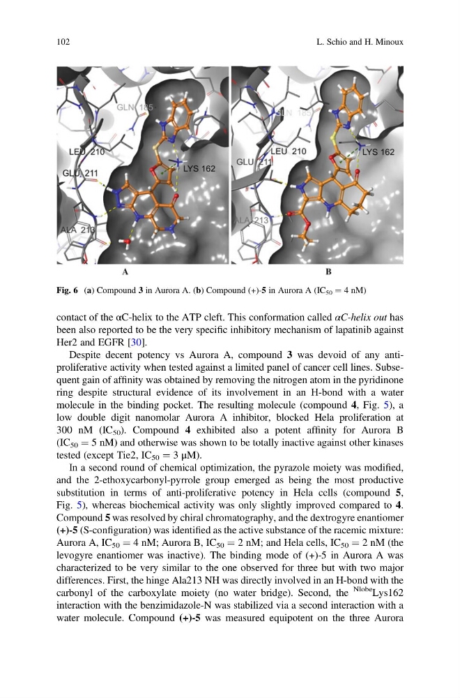
levogyre enantiomer was inactive). The binding mode of (+)-5 in Aurora A was
characterized to be very similar to the one observed for three but with two major
differences. First, the hinge Ala213 NH was directly involved in an H-bond with the
carbonyl of the carboxylate moiety (no water bridge). Second, the
Nlobe Lys162
interaction with the benzimidazole-N was stabilized via a second interaction with a
water molecule. Compound (+)-5 was measured equipotent on the three Aurora
Fig. 6 (a) Compound 3 in Aurora A. (b) Compound (+)-5 in Aurora A (IC 50 ¼ 4 nM)
102
L. Schio and H. Minoux
been also reported to be the very specific inhibitory mechanism of lapatinib against
Her2 and EGFR [30].
Despite decent potency vs Aurora A, compound 3 was devoid of any antiproliferative activity when tested against a limited panel of cancer cell lines. Subsequent gain of affinity was obtained by removing the nitrogen atom in the pyridinone
ring despite structural evidence of its involvement in an H-bond with a water
molecule in the binding pocket. The resulting molecule (compound 4, Fig. 5), a
low double digit nanomolar Aurora A inhibitor, blocked Hela proliferation at
300 nM (IC 50 ). Compound 4 exhibited also a potent affinity for Aurora B
(IC 50 ¼ 5 nM) and otherwise was shown to be totally inactive against other kinases
tested (except Tie2, IC 50 ¼ 3 μM).
In a second round of chemical optimization, the pyrazole moiety was modified,
and the 2-ethoxycarbonyl-pyrrole group emerged as being the most productive
substitution in terms of anti-proliferative potency in Hela cells (compound 5,
Fig. 5), whereas biochemical activity was only slightly improved compared to 4.
Compound 5 was resolved by chiral chromatography, and the dextrogyre enantiomer
(+)-5 (S-configuration) was identified as the active substance of the racemic mixture:
Aurora A, IC 50 ¼ 4 nM; Aurora B, IC 50 ¼ 2 nM; and Hela cells, IC 50 ¼ 2 nM (the
levogyre enantiomer was inactive). The binding mode of (+)-5 in Aurora A was
characterized to be very similar to the one observed for three but with two major
differences. First, the hinge Ala213 NH was directly involved in an H-bond with the
carbonyl of the carboxylate moiety (no water bridge). Second, the
Nlobe Lys162
interaction with the benzimidazole-N was stabilized via a second interaction with a
water molecule. Compound (+)-5 was measured equipotent on the three Aurora
Fig. 6 (a) Compound 3 in Aurora A. (b) Compound (+)-5 in Aurora A (IC 50 ¼ 4 nM)
102
L. Schio and H. Minoux