

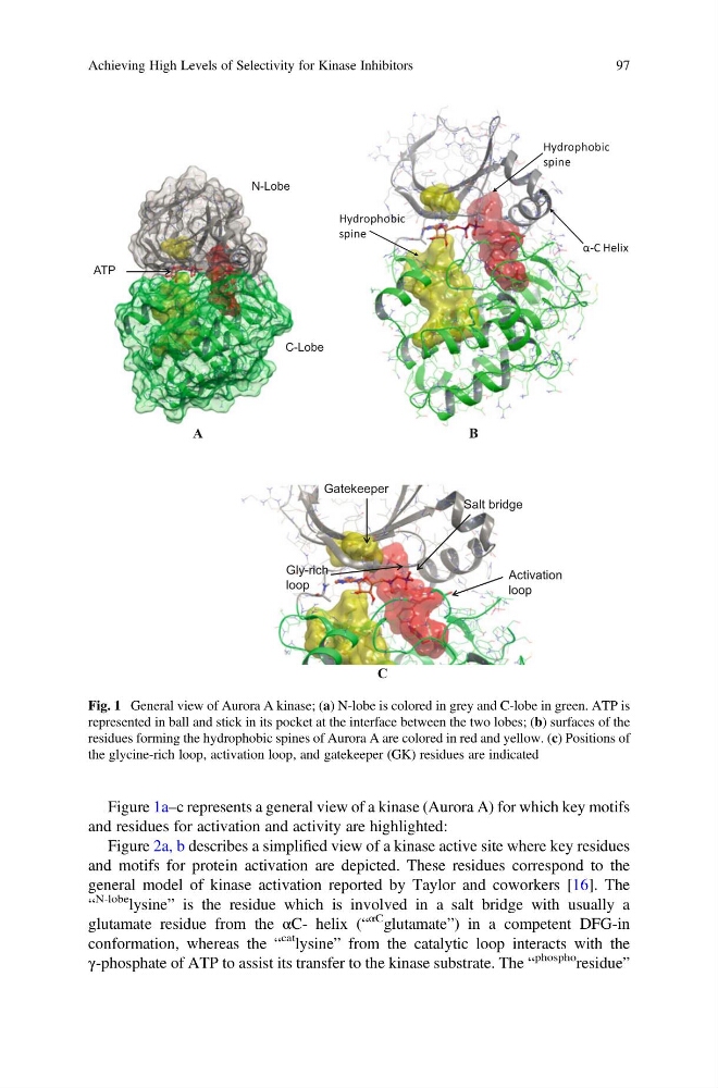
Figure 1a–c represents a general view of a kinase (Aurora A) for which key motifs
and residues for activation and activity are highlighted:
Figure 2a, b describes a simplified view of a kinase active site where key residues
and motifs for protein activation are depicted. These residues correspond to the
general model of kinase activation reported by Taylor and coworkers [16]. The
“
N-lobe lysine” is the residue which is involved in a salt bridge with usually a
glutamate residue from the αC- helix (“
αC glutamate”) in a competent DFG-in
conformation, whereas the “
cat lysine” from the catalytic loop interacts with the
γ-phosphate of ATP to assist its transfer to the kinase substrate. The “
phospho
residue”
A
B
C
N-Lobe
C-Lobe
ATP
α-C Helix
Hydrophobic
spine
Hydrophobic
spine
Gatekeeper
Activation
loop
Gly-rich
loop
Salt bridge
Fig. 1 General view of Aurora A kinase; (a) N-lobe is colored in grey and C-lobe in green. ATP is
represented in ball and stick in its pocket at the interface between the two lobes; (b) surfaces of the
residues forming the hydrophobic spines of Aurora A are colored in red and yellow. (c) Positions of
the glycine-rich loop, activation loop, and gatekeeper (GK) residues are indicated
Achieving High Levels of Selectivity for Kinase Inhibitors
97
and residues for activation and activity are highlighted:
Figure 2a, b describes a simplified view of a kinase active site where key residues
and motifs for protein activation are depicted. These residues correspond to the
general model of kinase activation reported by Taylor and coworkers [16]. The
“
N-lobe lysine” is the residue which is involved in a salt bridge with usually a
glutamate residue from the αC- helix (“
αC glutamate”) in a competent DFG-in
conformation, whereas the “
cat lysine” from the catalytic loop interacts with the
γ-phosphate of ATP to assist its transfer to the kinase substrate. The “
phospho
residue”
A
B
C
N-Lobe
C-Lobe
ATP
α-C Helix
Hydrophobic
spine
Hydrophobic
spine
Gatekeeper
Activation
loop
Gly-rich
loop
Salt bridge
Fig. 1 General view of Aurora A kinase; (a) N-lobe is colored in grey and C-lobe in green. ATP is
represented in ball and stick in its pocket at the interface between the two lobes; (b) surfaces of the
residues forming the hydrophobic spines of Aurora A are colored in red and yellow. (c) Positions of
the glycine-rich loop, activation loop, and gatekeeper (GK) residues are indicated
Achieving High Levels of Selectivity for Kinase Inhibitors
97