

10 Analytical Protocols in the Measurement …
233
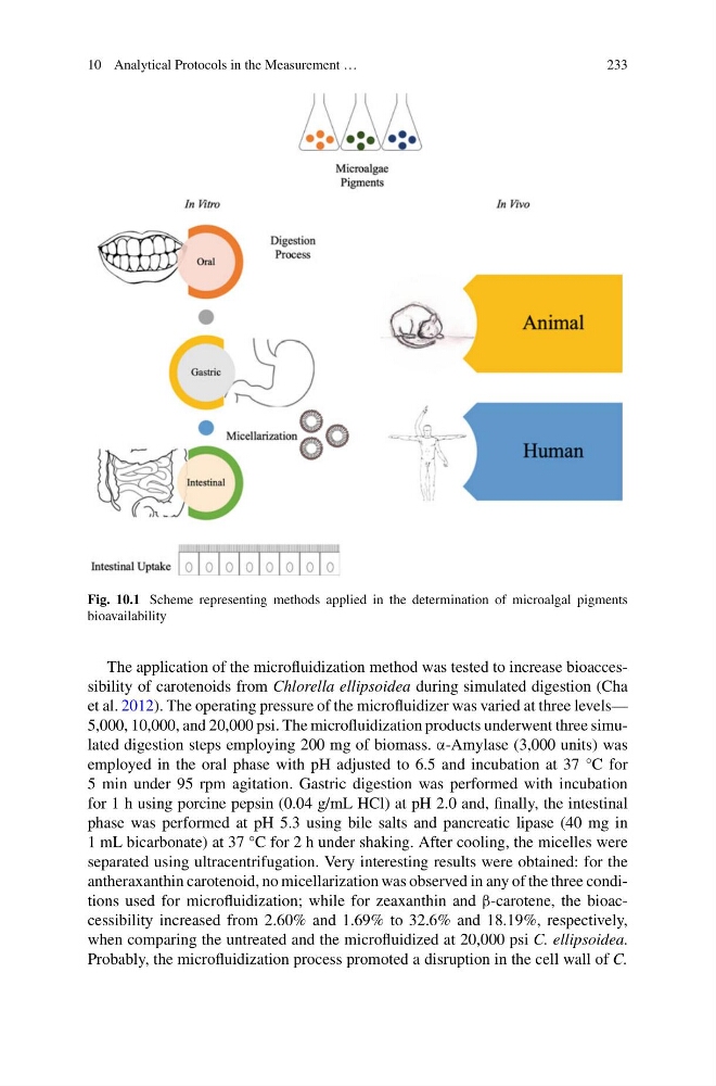
Fig. 10.1 Scheme representing methods applied in the determination of microalgal pigments
bioavailability
The application of the microfluidization method was tested to increase bioaccessibility of carotenoids from Chlorella ellipsoidea during simulated digestion (Cha
et al. 2012). The operating pressure of the microfluidizer was varied at three levels—
5,000, 10,000, and 20,000 psi. The microfluidization products underwent three simulated digestion steps employing 200 mg of biomass. α-Amylase (3,000 units) was
employed in the oral phase with pH adjusted to 6.5 and incubation at 37 °C for
5 min under 95 rpm agitation. Gastric digestion was performed with incubation
for 1 h using porcine pepsin (0.04 g/mL HCl) at pH 2.0 and, finally, the intestinal
phase was performed at pH 5.3 using bile salts and pancreatic lipase (40 mg in
1 mL bicarbonate) at 37 °C for 2 h under shaking. After cooling, the micelles were
separated using ultracentrifugation. Very interesting results were obtained: for the
antheraxanthin carotenoid, no micellarization was observed in any of the three conditions used for microfluidization; while for zeaxanthin and β-carotene, the bioaccessibility increased from 2.60% and 1.69% to 32.6% and 18.19%, respectively,
when comparing the untreated and the microfluidized at 20,000 psi C. ellipsoidea.
Probably, the microfluidization process promoted a disruption in the cell wall of C.
233
Fig. 10.1 Scheme representing methods applied in the determination of microalgal pigments
bioavailability
The application of the microfluidization method was tested to increase bioaccessibility of carotenoids from Chlorella ellipsoidea during simulated digestion (Cha
et al. 2012). The operating pressure of the microfluidizer was varied at three levels—
5,000, 10,000, and 20,000 psi. The microfluidization products underwent three simulated digestion steps employing 200 mg of biomass. α-Amylase (3,000 units) was
employed in the oral phase with pH adjusted to 6.5 and incubation at 37 °C for
5 min under 95 rpm agitation. Gastric digestion was performed with incubation
for 1 h using porcine pepsin (0.04 g/mL HCl) at pH 2.0 and, finally, the intestinal
phase was performed at pH 5.3 using bile salts and pancreatic lipase (40 mg in
1 mL bicarbonate) at 37 °C for 2 h under shaking. After cooling, the micelles were
separated using ultracentrifugation. Very interesting results were obtained: for the
antheraxanthin carotenoid, no micellarization was observed in any of the three conditions used for microfluidization; while for zeaxanthin and β-carotene, the bioaccessibility increased from 2.60% and 1.69% to 32.6% and 18.19%, respectively,
when comparing the untreated and the microfluidized at 20,000 psi C. ellipsoidea.
Probably, the microfluidization process promoted a disruption in the cell wall of C.