

8 Analytical Protocols in Phycobiliproteins Analysis
191
with lectins, such as jacalin (Pandey et al. 2009a), concanavalin A and peanut agglutinin (Pandey et al. 2009b), and PCB interactions with human serum albumin (HSA)
(Minic et al. 2015), bovine serum albumin (BSA) (Kathiravan et al. 2009; Minic
et al. 2018a), and beta-lactoglobulin (BLG) (Minic et al. 2018b). For identification
of PCB binding site on HSA, competition experiments with site markers, as well as
the determination of binding constants in the presence of site markers were exploited
(Minic et al. 2015). Fluorescence microscopy has shown that PC penetrates into
nucleated cells and stains the nucleus. High affinity of PC for DNA was confirmed
by agarose gel electrophoresis, suggesting that PC can be used as a natural non-toxic
replacement for ethidium bromide for specific detection of genomic DNA and as
a marker of various blood cells/molecules (Singh et al. 2011; Paswan et al. 2016).
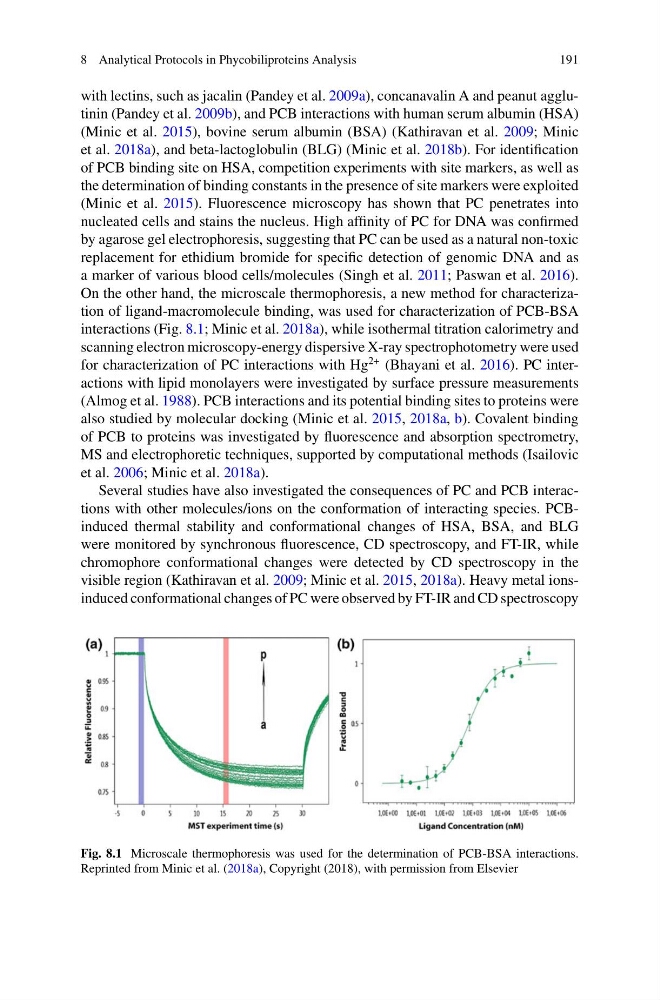
On the other hand, the microscale thermophoresis, a new method for characterization of ligand-macromolecule binding, was used for characterization of PCB-BSA
interactions (Fig. 8.1; Minic et al. 2018a), while isothermal titration calorimetry and
scanning electron microscopy-energy dispersive X-ray spectrophotometry were used
for characterization of PC interactions with Hg
2+ (Bhayani et al. 2016). PC interactions with lipid monolayers were investigated by surface pressure measurements
(Almog et al. 1988). PCB interactions and its potential binding sites to proteins were
also studied by molecular docking (Minic et al. 2015, 2018a, b). Covalent binding
of PCB to proteins was investigated by fluorescence and absorption spectrometry,
MS and electrophoretic techniques, supported by computational methods (Isailovic
et al. 2006; Minic et al. 2018a).
Several studies have also investigated the consequences of PC and PCB interactions with other molecules/ions on the conformation of interacting species. PCBinduced thermal stability and conformational changes of HSA, BSA, and BLG
were monitored by synchronous fluorescence, CD spectroscopy, and FT-IR, while
chromophore conformational changes were detected by CD spectroscopy in the
visible region (Kathiravan et al. 2009; Minic et al. 2015, 2018a). Heavy metal ionsinduced conformational changes of PC were observed by FT-IR and CD spectroscopy
Fig. 8.1 Microscale thermophoresis was used for the determination of PCB-BSA interactions.
Reprinted from Minic et al. (2018a), Copyright (2018), with permission from Elsevier
191
with lectins, such as jacalin (Pandey et al. 2009a), concanavalin A and peanut agglutinin (Pandey et al. 2009b), and PCB interactions with human serum albumin (HSA)
(Minic et al. 2015), bovine serum albumin (BSA) (Kathiravan et al. 2009; Minic
et al. 2018a), and beta-lactoglobulin (BLG) (Minic et al. 2018b). For identification
of PCB binding site on HSA, competition experiments with site markers, as well as
the determination of binding constants in the presence of site markers were exploited
(Minic et al. 2015). Fluorescence microscopy has shown that PC penetrates into
nucleated cells and stains the nucleus. High affinity of PC for DNA was confirmed
by agarose gel electrophoresis, suggesting that PC can be used as a natural non-toxic
replacement for ethidium bromide for specific detection of genomic DNA and as
a marker of various blood cells/molecules (Singh et al. 2011; Paswan et al. 2016).
On the other hand, the microscale thermophoresis, a new method for characterization of ligand-macromolecule binding, was used for characterization of PCB-BSA
interactions (Fig. 8.1; Minic et al. 2018a), while isothermal titration calorimetry and
scanning electron microscopy-energy dispersive X-ray spectrophotometry were used
for characterization of PC interactions with Hg
2+ (Bhayani et al. 2016). PC interactions with lipid monolayers were investigated by surface pressure measurements
(Almog et al. 1988). PCB interactions and its potential binding sites to proteins were
also studied by molecular docking (Minic et al. 2015, 2018a, b). Covalent binding
of PCB to proteins was investigated by fluorescence and absorption spectrometry,
MS and electrophoretic techniques, supported by computational methods (Isailovic
et al. 2006; Minic et al. 2018a).
Several studies have also investigated the consequences of PC and PCB interactions with other molecules/ions on the conformation of interacting species. PCBinduced thermal stability and conformational changes of HSA, BSA, and BLG
were monitored by synchronous fluorescence, CD spectroscopy, and FT-IR, while
chromophore conformational changes were detected by CD spectroscopy in the
visible region (Kathiravan et al. 2009; Minic et al. 2015, 2018a). Heavy metal ionsinduced conformational changes of PC were observed by FT-IR and CD spectroscopy
Fig. 8.1 Microscale thermophoresis was used for the determination of PCB-BSA interactions.
Reprinted from Minic et al. (2018a), Copyright (2018), with permission from Elsevier