

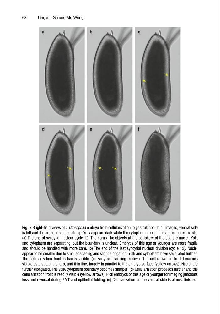
Fig. 2 Bright-field views of a Drosophila embryo from cellularization to gastrulation. In all images, ventral side
is left and the anterior side points up. Yolk appears dark while the cytoplasm appears as a transparent circle.
(a) The end of syncytial nuclear cycle 12. The bump-like objects at the periphery of the egg are nuclei. Yolk
and cytoplasm are separating, but the boundary is unclear. Embryos of this age or younger are more fragile
and should be handled with more care. (b) The end of the last syncytial nuclear division (cycle 13). Nuclei
appear to be smaller due to smaller spacing and slight elongation. Yolk and cytoplasm have separated further.
The cellularization front is hardly visible. (c) Early cellularizing embryo. The cellularization front becomes
visible as a straight, sharp, and thin line, largely in parallel to the embryo surface (yellow arrows). Nuclei are
further elongated. The yolk/cytoplasm boundary becomes sharper. (d) Cellularization proceeds further and the
cellularization front is readily visible (yellow arrows). Pick embryos of this age or younger for imaging junctions
loss and reversal during EMT and epithelial folding. (e) Cellularization on the ventral side is almost finished.
68
Lingkun Gu and Mo Weng
is left and the anterior side points up. Yolk appears dark while the cytoplasm appears as a transparent circle.
(a) The end of syncytial nuclear cycle 12. The bump-like objects at the periphery of the egg are nuclei. Yolk
and cytoplasm are separating, but the boundary is unclear. Embryos of this age or younger are more fragile
and should be handled with more care. (b) The end of the last syncytial nuclear division (cycle 13). Nuclei
appear to be smaller due to smaller spacing and slight elongation. Yolk and cytoplasm have separated further.
The cellularization front is hardly visible. (c) Early cellularizing embryo. The cellularization front becomes
visible as a straight, sharp, and thin line, largely in parallel to the embryo surface (yellow arrows). Nuclei are
further elongated. The yolk/cytoplasm boundary becomes sharper. (d) Cellularization proceeds further and the
cellularization front is readily visible (yellow arrows). Pick embryos of this age or younger for imaging junctions
loss and reversal during EMT and epithelial folding. (e) Cellularization on the ventral side is almost finished.
68
Lingkun Gu and Mo Weng