

following cellularization, gastrulation starts with the epithelial folding of the mesoderm to form the ventral furrow. This ventral
furrow formation is driven by mechanical force generated from
myosin contraction, on the apical surface of the mesoderm. This
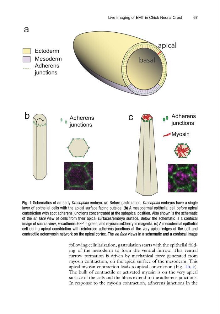
apical myosin contraction leads to apical constriction (Fig. 1b, c).
The bulk of contractile or activated myosin is on the very apical
surface of the cells and the fibers extend to the adherens junctions.
In response to the myosin contraction, adherens junctions in the
Fig. 1 Schematics of an early Drosophila embryo. (a) Before gastrulation, Drosophila embryos have a single
layer of epithelial cells with the apical surface facing outside. (b) A mesodermal epithelial cell before apical
constriction with spot adherens junctions concentrated at the subapical position. Also shown is the schematic
of the en face view of cells from their apical surfaces/embryo surface. Below the schematic is a confocal
image of such a view, E-cadherin::GFP in green, and myosin::mCherry in magenta. (c) A mesodermal epithelial
cell during apical constriction with reinforced adherens junctions at the very apical edges of the cell and
contractile actomyosin network on the apical cortex. The en face views in a schematic and a confocal image
Live Imaging of EMT in Chick Neural Crest
67
furrow formation is driven by mechanical force generated from
myosin contraction, on the apical surface of the mesoderm. This
apical myosin contraction leads to apical constriction (Fig. 1b, c).
The bulk of contractile or activated myosin is on the very apical
surface of the cells and the fibers extend to the adherens junctions.
In response to the myosin contraction, adherens junctions in the
Fig. 1 Schematics of an early Drosophila embryo. (a) Before gastrulation, Drosophila embryos have a single
layer of epithelial cells with the apical surface facing outside. (b) A mesodermal epithelial cell before apical
constriction with spot adherens junctions concentrated at the subapical position. Also shown is the schematic
of the en face view of cells from their apical surfaces/embryo surface. Below the schematic is a confocal
image of such a view, E-cadherin::GFP in green, and myosin::mCherry in magenta. (c) A mesodermal epithelial
cell during apical constriction with reinforced adherens junctions at the very apical edges of the cell and
contractile actomyosin network on the apical cortex. The en face views in a schematic and a confocal image
Live Imaging of EMT in Chick Neural Crest
67