

Ct (GOI,T) being the Ct of the Gene Of Interest (GOI, also
called “target” gene) in the test sample (i.e., TGF-β -treated (T) condition).
Ct (GOI,NT) being the Ct of the GOI in the calibrator sample
(i.e., non-treated (NT) condition).
Ct (RG,T) being the Ct of the Reference Gene (RG) in the T
condition.
Ct (RG,NT) being the Ct of the RG in the NT condition.
In detail:
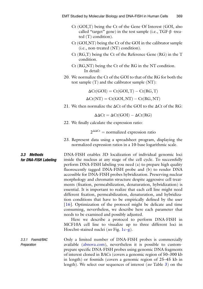
20. We normalize the Ct of the GOI to that of the RG for both the
test sample (T) and the calibrator sample (NT):
ΔCt GOI
ð
Þ¼Ct GOI, T
ð
ÞÀCt RG, T
ð
Þ
ΔCt NT
ð
Þ ¼ Ct GOI, NT
ð
ÞÀCt RG, NT
ð
Þ
21. We then normalize the ΔCt of the GOI to the ΔCt of the RG:
ΔΔCt ¼ ΔCt GOI
ð
ÞÀΔCt RG
ð Þ
22. We finally calculate the expression ratio:
2
ΔΔCt
¼ normalized expression ratio
23. Represent data using a spreadsheet program, displaying the
normalized expression ratios in a 10-base logarithmic scale.
3.3 Methods
for DNA-FISH Labeling
DNA-FISH enables 3D localization of individual genomic loci
inside the nucleus at any stage of the cell cycle. To successfully
perform DNA-FISH labeling you need (a) to prepare high quality
fluorescently tagged DNA-FISH probe and (b) to render DNA
accessible for DNA-FISH probes hybridization. Preserving nuclear
morphology and chromatin structure despite aggressive cell treatments (fixation, permeabilization, denaturation, hybridization) is
essential. It is important to realize that each cell line might need
different fixation, permeabilization, denaturation, and hybridization conditions that have to be empirically defined by the user
[16]. Optimization of the protocol might be delicate and time
consuming, nevertheless, we describe here each parameter that
needs to be examined and possibly adjusted.
Here we describe a protocol to perform DNA-FISH in
MCF10A cell line to visualize up to three different loci in
Hoechst-stained nuclei (see Fig. 1c–g).
3.3.1 Fosmid/BAC
Preparation
Only a limited number of DNA-FISH probes is commercially
available (abnova.com), nevertheless it is possible to customprepare specific DNA-FISH probes using genomic DNA fragments
of interest cloned in BACs (covers a genomic region of 50–300 kb
in length) or fosmids (covers a genomic region of 25–45 kb in
length). We select our sequences of interest (see Table 3) on the
EMT Studied by Molecular Biology and DNA-FISH in Human Cells
369
called “target” gene) in the test sample (i.e., TGF-β -treated (T) condition).
Ct (GOI,NT) being the Ct of the GOI in the calibrator sample
(i.e., non-treated (NT) condition).
Ct (RG,T) being the Ct of the Reference Gene (RG) in the T
condition.
Ct (RG,NT) being the Ct of the RG in the NT condition.
In detail:
20. We normalize the Ct of the GOI to that of the RG for both the
test sample (T) and the calibrator sample (NT):
ΔCt GOI
ð
Þ¼Ct GOI, T
ð
ÞÀCt RG, T
ð
Þ
ΔCt NT
ð
Þ ¼ Ct GOI, NT
ð
ÞÀCt RG, NT
ð
Þ
21. We then normalize the ΔCt of the GOI to the ΔCt of the RG:
ΔΔCt ¼ ΔCt GOI
ð
ÞÀΔCt RG
ð Þ
22. We finally calculate the expression ratio:
2
ΔΔCt
¼ normalized expression ratio
23. Represent data using a spreadsheet program, displaying the
normalized expression ratios in a 10-base logarithmic scale.
3.3 Methods
for DNA-FISH Labeling
DNA-FISH enables 3D localization of individual genomic loci
inside the nucleus at any stage of the cell cycle. To successfully
perform DNA-FISH labeling you need (a) to prepare high quality
fluorescently tagged DNA-FISH probe and (b) to render DNA
accessible for DNA-FISH probes hybridization. Preserving nuclear
morphology and chromatin structure despite aggressive cell treatments (fixation, permeabilization, denaturation, hybridization) is
essential. It is important to realize that each cell line might need
different fixation, permeabilization, denaturation, and hybridization conditions that have to be empirically defined by the user
[16]. Optimization of the protocol might be delicate and time
consuming, nevertheless, we describe here each parameter that
needs to be examined and possibly adjusted.
Here we describe a protocol to perform DNA-FISH in
MCF10A cell line to visualize up to three different loci in
Hoechst-stained nuclei (see Fig. 1c–g).
3.3.1 Fosmid/BAC
Preparation
Only a limited number of DNA-FISH probes is commercially
available (abnova.com), nevertheless it is possible to customprepare specific DNA-FISH probes using genomic DNA fragments
of interest cloned in BACs (covers a genomic region of 50–300 kb
in length) or fosmids (covers a genomic region of 25–45 kb in
length). We select our sequences of interest (see Table 3) on the
EMT Studied by Molecular Biology and DNA-FISH in Human Cells
369