

Here, we present a protocol to perform MTM of intercellular
adhesion proteins E-cadherins [13] that experience cytoskeletongenerated tension sensitive to external cues [14]. During
Epithelial-Mesenchymal Transition (EMT), E-cadherin tension
relaxes, which associates with the release of its interactant
β-catenin and activation of β-catenin dependent transcription [15].
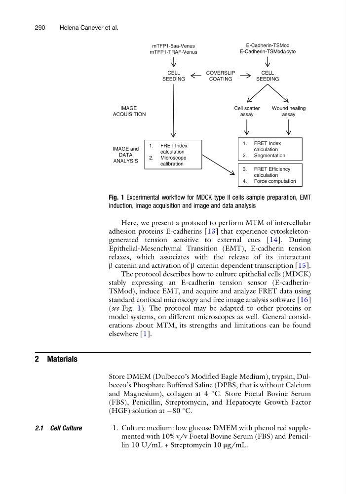
The protocol describes how to culture epithelial cells (MDCK)
stably expressing an E-cadherin tension sensor (E-cadherinTSMod), induce EMT, and acquire and analyze FRET data using
standard confocal microscopy and free image analysis software [16]
(see Fig. 1). The protocol may be adapted to other proteins or
model systems, on different microscopes as well. General considerations about MTM, its strengths and limitations can be found
elsewhere [1].
2 Materials
Store DMEM (Dulbecco’s Modified Eagle Medium), trypsin, Dulbecco’s Phosphate Buffered Saline (DPBS, that is without Calcium
and Magnesium), collagen at 4
C. Store Foetal Bovine Serum
(FBS), Penicillin, Streptomycin, and Hepatocyte Growth Factor
(HGF) solution at À80
C.
2.1 Cell Culture
1. Culture medium: low glucose DMEM with phenol red supplemented with 10% v/v Foetal Bovine Serum (FBS) and Penicillin 10 U/mL + Streptomycin 10 μg/mL.
CELL
SEEDING
Cell scatter
assay
Wound healing
assay
IMAGE and
DATA
ANALYSIS
mTFP1-5aa-Venus
mTFP1-TRAF-Venus
E-Cadherin-TSMod
E-Cadherin-TSModΔcyto
COVERSLIP
COATING
IMAGE
ACQUISITION
CELL
SEEDING
1. FRET Index
calculation
2. Microscope
calibration
1. FRET Index
calculation
2. Segmentation
3. FRET Efficiency
calculation
4. Force computation
Fig. 1 Experimental workflow for MDCK type II cells sample preparation, EMT
induction, image acquisition and image and data analysis
290
Helena Canever et al.
adhesion proteins E-cadherins [13] that experience cytoskeletongenerated tension sensitive to external cues [14]. During
Epithelial-Mesenchymal Transition (EMT), E-cadherin tension
relaxes, which associates with the release of its interactant
β-catenin and activation of β-catenin dependent transcription [15].
The protocol describes how to culture epithelial cells (MDCK)
stably expressing an E-cadherin tension sensor (E-cadherinTSMod), induce EMT, and acquire and analyze FRET data using
standard confocal microscopy and free image analysis software [16]
(see Fig. 1). The protocol may be adapted to other proteins or
model systems, on different microscopes as well. General considerations about MTM, its strengths and limitations can be found
elsewhere [1].
2 Materials
Store DMEM (Dulbecco’s Modified Eagle Medium), trypsin, Dulbecco’s Phosphate Buffered Saline (DPBS, that is without Calcium
and Magnesium), collagen at 4
C. Store Foetal Bovine Serum
(FBS), Penicillin, Streptomycin, and Hepatocyte Growth Factor
(HGF) solution at À80
C.
2.1 Cell Culture
1. Culture medium: low glucose DMEM with phenol red supplemented with 10% v/v Foetal Bovine Serum (FBS) and Penicillin 10 U/mL + Streptomycin 10 μg/mL.
CELL
SEEDING
Cell scatter
assay
Wound healing
assay
IMAGE and
DATA
ANALYSIS
mTFP1-5aa-Venus
mTFP1-TRAF-Venus
E-Cadherin-TSMod
E-Cadherin-TSModΔcyto
COVERSLIP
COATING
IMAGE
ACQUISITION
CELL
SEEDING
1. FRET Index
calculation
2. Microscope
calibration
1. FRET Index
calculation
2. Segmentation
3. FRET Efficiency
calculation
4. Force computation
Fig. 1 Experimental workflow for MDCK type II cells sample preparation, EMT
induction, image acquisition and image and data analysis
290
Helena Canever et al.