

19. Typically, 458, 477, 488, or 514 nm lasers are used for green
fluorescent proteins; 543, 561, and 568 nm for red; and 633 or
647 for far red.
20. If imaging a sample with more than one fluorescent marker, use
a dichromatic beamsplitter to acquire multiple wavelengths
without having to alter the illumination path, thus optimizing
the temporal resolution of the acquisition.
21. Be mindful of laser power and exposure time, as excessive laser
exposure can cause photobleaching (loss of fluorescence) and
phototoxicity (cell damage) to the sample.
22. Set laser power and exposure time using the image histogram
as a reference. Ideally, the histogram of the images should fill
the dynamic range of the camera with a minimal number of
saturated or underexposed pixels (aim for less than 5% in each
case) (Fig. 3).
23. Finer Z and temporal resolutions can provide valuable information about cell and protein dynamics, but they also increase
photobleaching. Depending on the fluorescent protein, imaging tissue slices 10-μm-thick, at 0.5 μm intervals every 15–30 s,
can provide relatively high Z and temporal resolutions with
minimal photobleaching for imaging sessions with durations
of approximately 1 h.
24. Typically, using a Micropoint laser with the neutral density
filter in positions 20–25, and the mechanical attenuator
between 50–90% transmittance, ten pulses on a single spot
can be used to sever a junction in approximately 670 ms. If
rapid laser ablation is required, use fewer pulses at a higher laser
power.
25. Laser ablation of Drosophila embryos using ultraviolet light
causes photoactivation of the vitelline membrane.
26. If wounding along a line or within a shape, it will be necessary
to create discrete spots representing those geometries. Metamorph has an option (“segment region”) to break any shape
into discrete spots with specific inter-spot distances.
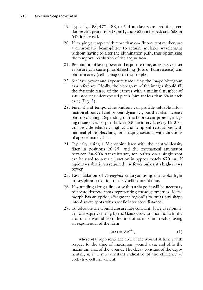
27. To calculate the wound closure rate constant, k, we use nonlinear least-squares fitting by the Gauss-Newton method to fit the
area of the wound from the time of its maximum value, using
an exponential of the form:
a t
ð Þ ¼ Ae
Àkt ,
ð1Þ
where a(t) represents the area of the wound at time t with
respect to the time of maximum wound area, and A is the
maximum area of the wound. The decay constant of the exponential, k, is a rate constant indicative of the efficiency of
collective cell movement.
216
Gordana Scepanovic et al.
fluorescent proteins; 543, 561, and 568 nm for red; and 633 or
647 for far red.
20. If imaging a sample with more than one fluorescent marker, use
a dichromatic beamsplitter to acquire multiple wavelengths
without having to alter the illumination path, thus optimizing
the temporal resolution of the acquisition.
21. Be mindful of laser power and exposure time, as excessive laser
exposure can cause photobleaching (loss of fluorescence) and
phototoxicity (cell damage) to the sample.
22. Set laser power and exposure time using the image histogram
as a reference. Ideally, the histogram of the images should fill
the dynamic range of the camera with a minimal number of
saturated or underexposed pixels (aim for less than 5% in each
case) (Fig. 3).
23. Finer Z and temporal resolutions can provide valuable information about cell and protein dynamics, but they also increase
photobleaching. Depending on the fluorescent protein, imaging tissue slices 10-μm-thick, at 0.5 μm intervals every 15–30 s,
can provide relatively high Z and temporal resolutions with
minimal photobleaching for imaging sessions with durations
of approximately 1 h.
24. Typically, using a Micropoint laser with the neutral density
filter in positions 20–25, and the mechanical attenuator
between 50–90% transmittance, ten pulses on a single spot
can be used to sever a junction in approximately 670 ms. If
rapid laser ablation is required, use fewer pulses at a higher laser
power.
25. Laser ablation of Drosophila embryos using ultraviolet light
causes photoactivation of the vitelline membrane.
26. If wounding along a line or within a shape, it will be necessary
to create discrete spots representing those geometries. Metamorph has an option (“segment region”) to break any shape
into discrete spots with specific inter-spot distances.
27. To calculate the wound closure rate constant, k, we use nonlinear least-squares fitting by the Gauss-Newton method to fit the
area of the wound from the time of its maximum value, using
an exponential of the form:
a t
ð Þ ¼ Ae
Àkt ,
ð1Þ
where a(t) represents the area of the wound at time t with
respect to the time of maximum wound area, and A is the
maximum area of the wound. The decay constant of the exponential, k, is a rate constant indicative of the efficiency of
collective cell movement.
216
Gordana Scepanovic et al.