

9. A slide to break the tip of microneedles (see Note 4).
10. Micromanipulator.
11. Pneumatic pump.
12. Inverted microscope with transmitted light illumination and a
10Â or 20Â air objective, coupled to the micromanipulator
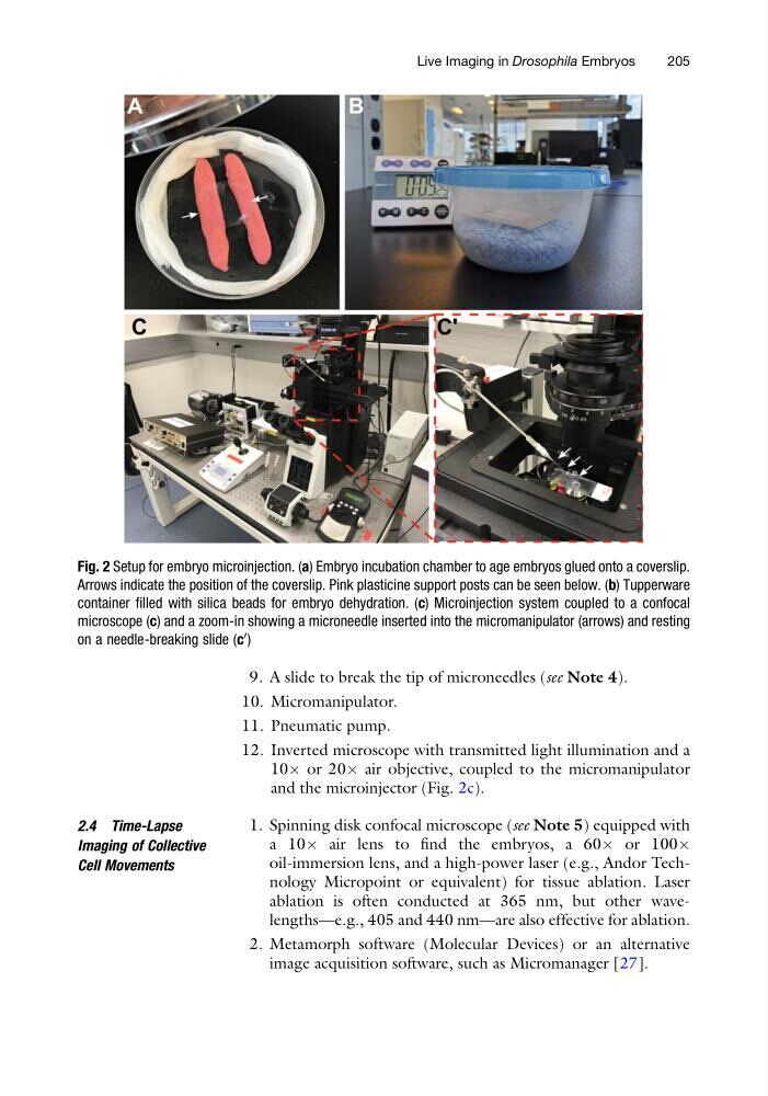
and the microinjector (Fig. 2c).
2.4 Time-Lapse
Imaging of Collective
Cell Movements
1. Spinning disk confocal microscope (see Note 5) equipped with
a 10Â air lens to find the embryos, a 60Â or 100Â
oil-immersion lens, and a high-power laser (e.g., Andor Technology Micropoint or equivalent) for tissue ablation. Laser
ablation is often conducted at 365 nm, but other wavelengths—e.g., 405 and 440 nm—are also effective for ablation.
2. Metamorph software (Molecular Devices) or an alternative
image acquisition software, such as Micromanager [27].
Fig. 2 Setup for embryo microinjection. (a) Embryo incubation chamber to age embryos glued onto a coverslip.
Arrows indicate the position of the coverslip. Pink plasticine support posts can be seen below. (b) Tupperware
container filled with silica beads for embryo dehydration. (c) Microinjection system coupled to a confocal
microscope (c) and a zoom-in showing a microneedle inserted into the micromanipulator (arrows) and resting
on a needle-breaking slide (c
0 )
Live Imaging in Drosophila Embryos
205
10. Micromanipulator.
11. Pneumatic pump.
12. Inverted microscope with transmitted light illumination and a
10Â or 20Â air objective, coupled to the micromanipulator
and the microinjector (Fig. 2c).
2.4 Time-Lapse
Imaging of Collective
Cell Movements
1. Spinning disk confocal microscope (see Note 5) equipped with
a 10Â air lens to find the embryos, a 60Â or 100Â
oil-immersion lens, and a high-power laser (e.g., Andor Technology Micropoint or equivalent) for tissue ablation. Laser
ablation is often conducted at 365 nm, but other wavelengths—e.g., 405 and 440 nm—are also effective for ablation.
2. Metamorph software (Molecular Devices) or an alternative
image acquisition software, such as Micromanager [27].
Fig. 2 Setup for embryo microinjection. (a) Embryo incubation chamber to age embryos glued onto a coverslip.
Arrows indicate the position of the coverslip. Pink plasticine support posts can be seen below. (b) Tupperware
container filled with silica beads for embryo dehydration. (c) Microinjection system coupled to a confocal
microscope (c) and a zoom-in showing a microneedle inserted into the micromanipulator (arrows) and resting
on a needle-breaking slide (c
0 )
Live Imaging in Drosophila Embryos
205