

3.3 Detailed
Characterization
of Primary Tumor
Burden in Flies
1. For a detailed characterization of primary tumor burden, dissect the gastrointestinal tract from ten female mated flies at a
precise time point, e.g., 3 weeks after heat shock. Dissect off
the MpTs (see Note 4).
2. Fix the midguts (see Note 3) and immunostain for GFP to mark
the clones, and Phalloidin to demark the midgut.
3. Image a single z-plane of the entire midgut on a confocal
microscope with a 10Â lens (see Note 5). On average a female
midgut is around 0.4 cm long, therefore imaging in tile-scan
mode is the most efficient way of acquiring this data. If this is
not available, then image the entire midgut and stitch the
multiple images together on Fiji.
4. Import a single image of an entire midgut into Fiji and use the
freehand selection to draw around tumors. Use the measure
tool to measure the area of each clone. Add up the total area of
the midgut covered by GFP, divide by the total area of the gut
and multiply by 100. This will give the total GFP% coverage of
the midgut. In a second measurement which we find is a more
robust measure for tumors is to calculate the average size of
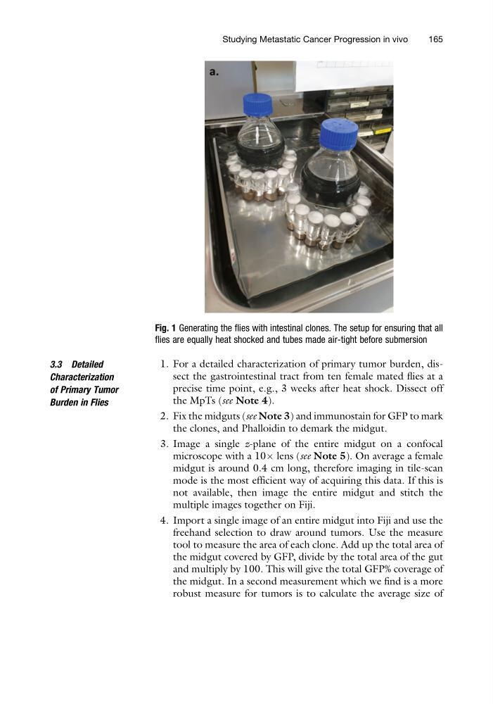
Fig. 1 Generating the flies with intestinal clones. The setup for ensuring that all
flies are equally heat shocked and tubes made air-tight before submersion
Studying Metastatic Cancer Progression in vivo
165
Characterization
of Primary Tumor
Burden in Flies
1. For a detailed characterization of primary tumor burden, dissect the gastrointestinal tract from ten female mated flies at a
precise time point, e.g., 3 weeks after heat shock. Dissect off
the MpTs (see Note 4).
2. Fix the midguts (see Note 3) and immunostain for GFP to mark
the clones, and Phalloidin to demark the midgut.
3. Image a single z-plane of the entire midgut on a confocal
microscope with a 10Â lens (see Note 5). On average a female
midgut is around 0.4 cm long, therefore imaging in tile-scan
mode is the most efficient way of acquiring this data. If this is
not available, then image the entire midgut and stitch the
multiple images together on Fiji.
4. Import a single image of an entire midgut into Fiji and use the
freehand selection to draw around tumors. Use the measure
tool to measure the area of each clone. Add up the total area of
the midgut covered by GFP, divide by the total area of the gut
and multiply by 100. This will give the total GFP% coverage of
the midgut. In a second measurement which we find is a more
robust measure for tumors is to calculate the average size of
Fig. 1 Generating the flies with intestinal clones. The setup for ensuring that all
flies are equally heat shocked and tubes made air-tight before submersion
Studying Metastatic Cancer Progression in vivo
165