

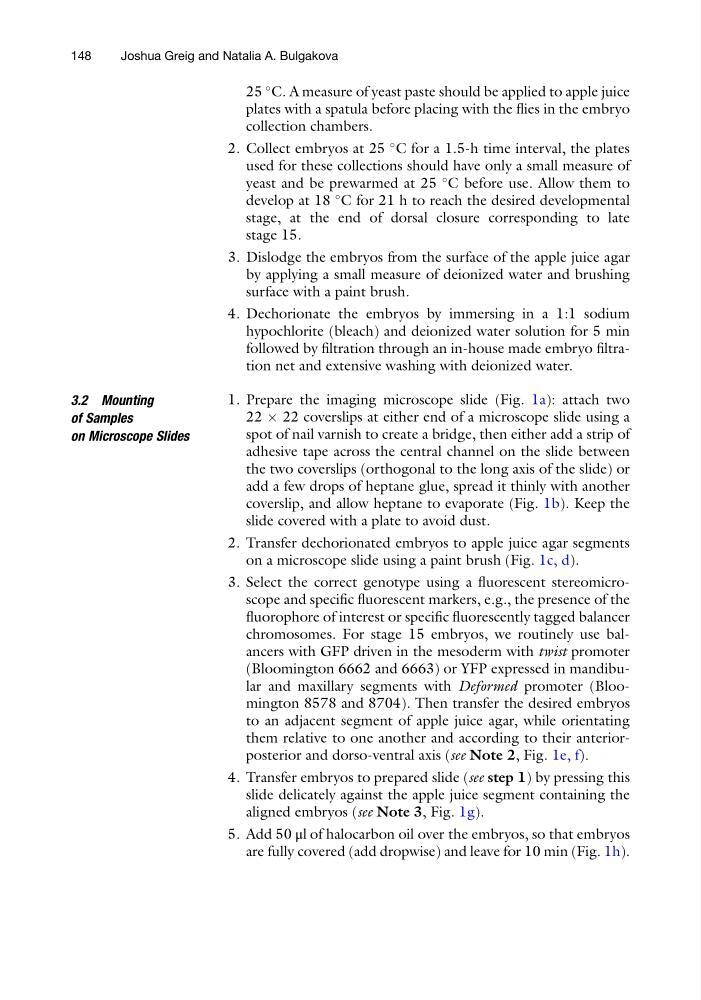
25
C. A measure of yeast paste should be applied to apple juice
plates with a spatula before placing with the flies in the embryo
collection chambers.
2. Collect embryos at 25
C for a 1.5-h time interval, the plates
used for these collections should have only a small measure of
yeast and be prewarmed at 25
C before use. Allow them to
develop at 18
C for 21 h to reach the desired developmental
stage, at the end of dorsal closure corresponding to late
stage 15.
3. Dislodge the embryos from the surface of the apple juice agar
by applying a small measure of deionized water and brushing
surface with a paint brush.
4. Dechorionate the embryos by immersing in a 1:1 sodium
hypochlorite (bleach) and deionized water solution for 5 min
followed by filtration through an in-house made embryo filtration net and extensive washing with deionized water.
3.2 Mounting
of Samples
on Microscope Slides
1. Prepare the imaging microscope slide (Fig. 1a): attach two
22 Â 22 coverslips at either end of a microscope slide using a
spot of nail varnish to create a bridge, then either add a strip of
adhesive tape across the central channel on the slide between
the two coverslips (orthogonal to the long axis of the slide) or
add a few drops of heptane glue, spread it thinly with another
coverslip, and allow heptane to evaporate (Fig. 1b). Keep the
slide covered with a plate to avoid dust.
2. Transfer dechorionated embryos to apple juice agar segments
on a microscope slide using a paint brush (Fig. 1c, d).
3. Select the correct genotype using a fluorescent stereomicroscope and specific fluorescent markers, e.g., the presence of the
fluorophore of interest or specific fluorescently tagged balancer
chromosomes. For stage 15 embryos, we routinely use balancers with GFP driven in the mesoderm with twist promoter
(Bloomington 6662 and 6663) or YFP expressed in mandibular and maxillary segments with Deformed promoter (Bloomington 8578 and 8704). Then transfer the desired embryos
to an adjacent segment of apple juice agar, while orientating
them relative to one another and according to their anteriorposterior and dorso-ventral axis (see Note 2, Fig. 1e, f).
4. Transfer embryos to prepared slide (see step 1) by pressing this
slide delicately against the apple juice segment containing the
aligned embryos (see Note 3, Fig. 1g).
5. Add 50 μl of halocarbon oil over the embryos, so that embryos
are fully covered (add dropwise) and leave for 10 min (Fig. 1h).
148
Joshua Greig and Natalia A. Bulgakova
C. A measure of yeast paste should be applied to apple juice
plates with a spatula before placing with the flies in the embryo
collection chambers.
2. Collect embryos at 25
C for a 1.5-h time interval, the plates
used for these collections should have only a small measure of
yeast and be prewarmed at 25
C before use. Allow them to
develop at 18
C for 21 h to reach the desired developmental
stage, at the end of dorsal closure corresponding to late
stage 15.
3. Dislodge the embryos from the surface of the apple juice agar
by applying a small measure of deionized water and brushing
surface with a paint brush.
4. Dechorionate the embryos by immersing in a 1:1 sodium
hypochlorite (bleach) and deionized water solution for 5 min
followed by filtration through an in-house made embryo filtration net and extensive washing with deionized water.
3.2 Mounting
of Samples
on Microscope Slides
1. Prepare the imaging microscope slide (Fig. 1a): attach two
22 Â 22 coverslips at either end of a microscope slide using a
spot of nail varnish to create a bridge, then either add a strip of
adhesive tape across the central channel on the slide between
the two coverslips (orthogonal to the long axis of the slide) or
add a few drops of heptane glue, spread it thinly with another
coverslip, and allow heptane to evaporate (Fig. 1b). Keep the
slide covered with a plate to avoid dust.
2. Transfer dechorionated embryos to apple juice agar segments
on a microscope slide using a paint brush (Fig. 1c, d).
3. Select the correct genotype using a fluorescent stereomicroscope and specific fluorescent markers, e.g., the presence of the
fluorophore of interest or specific fluorescently tagged balancer
chromosomes. For stage 15 embryos, we routinely use balancers with GFP driven in the mesoderm with twist promoter
(Bloomington 6662 and 6663) or YFP expressed in mandibular and maxillary segments with Deformed promoter (Bloomington 8578 and 8704). Then transfer the desired embryos
to an adjacent segment of apple juice agar, while orientating
them relative to one another and according to their anteriorposterior and dorso-ventral axis (see Note 2, Fig. 1e, f).
4. Transfer embryos to prepared slide (see step 1) by pressing this
slide delicately against the apple juice segment containing the
aligned embryos (see Note 3, Fig. 1g).
5. Add 50 μl of halocarbon oil over the embryos, so that embryos
are fully covered (add dropwise) and leave for 10 min (Fig. 1h).
148
Joshua Greig and Natalia A. Bulgakova