

3. Transfer wing discs to a new cavity block with ~250–500 μl
culture medium.
4. Place a 60 mm petri dish lid over the cavity block, place in a
humidified chamber, and move chamber to an incubator at the
desired temperature (29
C if using GAL80
ts
). Our chamber is
a simple plastic box with wet paper towel lining the base which
we enclose in saran wrap.
5. To analyze the onset of pEMT, culture discs for 7–8 h, and then
fix them in 4% formaldehyde in PBS for 15 min. Wash several
times in PBST and then continue with immunostaining using
standard methods.
A
B
C
D
F
E
DE-Cad
Viking-GFP
DE-Cad
DE-Cad
Viking-GFP
dp
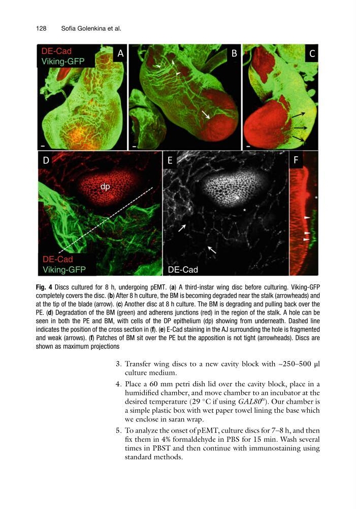
Fig. 4 Discs cultured for 8 h, undergoing pEMT. (a) A third-instar wing disc before culturing. Viking-GFP
completely covers the disc. (b) After 8 h culture, the BM is becoming degraded near the stalk (arrowheads) and
at the tip of the blade (arrow). (c) Another disc at 8 h culture. The BM is degrading and pulling back over the
PE. (d) Degradation of the BM (green) and adherens junctions (red) in the region of the stalk. A hole can be
seen in both the PE and BM, with cells of the DP epithelium (dp) showing from underneath. Dashed line
indicates the position of the cross section in (f). (e) E-Cad staining in the AJ surrounding the hole is fragmented
and weak (arrows). (f) Patches of BM sit over the PE but the apposition is not tight (arrowheads). Discs are
shown as maximum projections
128
Sofia Golenkina et al.
culture medium.
4. Place a 60 mm petri dish lid over the cavity block, place in a
humidified chamber, and move chamber to an incubator at the
desired temperature (29
C if using GAL80
ts
). Our chamber is
a simple plastic box with wet paper towel lining the base which
we enclose in saran wrap.
5. To analyze the onset of pEMT, culture discs for 7–8 h, and then
fix them in 4% formaldehyde in PBS for 15 min. Wash several
times in PBST and then continue with immunostaining using
standard methods.
A
B
C
D
F
E
DE-Cad
Viking-GFP
DE-Cad
DE-Cad
Viking-GFP
dp
Fig. 4 Discs cultured for 8 h, undergoing pEMT. (a) A third-instar wing disc before culturing. Viking-GFP
completely covers the disc. (b) After 8 h culture, the BM is becoming degraded near the stalk (arrowheads) and
at the tip of the blade (arrow). (c) Another disc at 8 h culture. The BM is degrading and pulling back over the
PE. (d) Degradation of the BM (green) and adherens junctions (red) in the region of the stalk. A hole can be
seen in both the PE and BM, with cells of the DP epithelium (dp) showing from underneath. Dashed line
indicates the position of the cross section in (f). (e) E-Cad staining in the AJ surrounding the hole is fragmented
and weak (arrows). (f) Patches of BM sit over the PE but the apposition is not tight (arrowheads). Discs are
shown as maximum projections
128
Sofia Golenkina et al.