

total) can be used according to the manufacturer’s instructions.
Briefly: Prepare five dilutions of a protein standard, which is
representative of the protein solution to be tested. The linear
range of the assay for bovine serum albumin (BSA) is
1.0–10.0 μg/mL. Prepare dye reagent by diluting 1 part Dye
Reagent Concentrate (Bio-Rad) with 4 parts of distilled water.
Mix well and keep at room temperature. Pipette 10 μL of each
BSA standard and sample solution into separate microtiter
plate wells (in triplicates). Add 200 μL of diluted dye reagent
to each well. Mix the sample and reagent thoroughly using a
microplate mixer. Incubate at room temperature for at least
5 min but no more than 1 h. Use 595 nm wavelength to
measure absorbance.
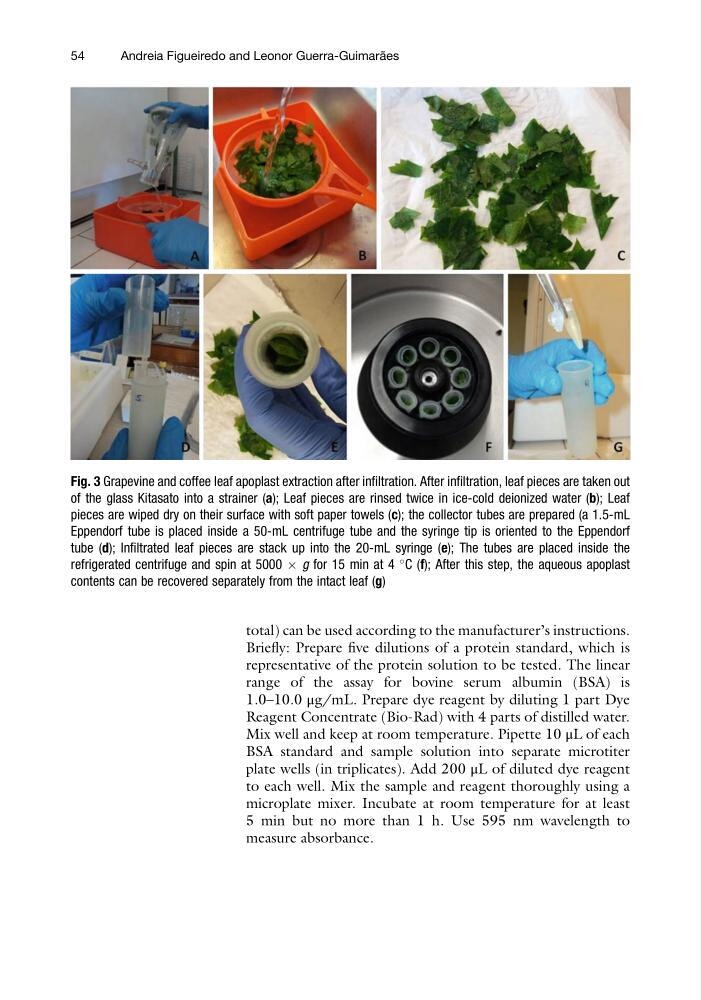
Fig. 3 Grapevine and coffee leaf apoplast extraction after infiltration. After infiltration, leaf pieces are taken out
of the glass Kitasato into a strainer (a); Leaf pieces are rinsed twice in ice-cold deionized water (b); Leaf
pieces are wiped dry on their surface with soft paper towels (c); the collector tubes are prepared (a 1.5-mL
Eppendorf tube is placed inside a 50-mL centrifuge tube and the syringe tip is oriented to the Eppendorf
tube (d); Infiltrated leaf pieces are stack up into the 20-mL syringe (e); The tubes are placed inside the
refrigerated centrifuge and spin at 5000 Â g for 15 min at 4
C (f); After this step, the aqueous apoplast
contents can be recovered separately from the intact leaf (g)
54
Andreia Figueiredo and Leonor Guerra-Guimara ˜ es
Briefly: Prepare five dilutions of a protein standard, which is
representative of the protein solution to be tested. The linear
range of the assay for bovine serum albumin (BSA) is
1.0–10.0 μg/mL. Prepare dye reagent by diluting 1 part Dye
Reagent Concentrate (Bio-Rad) with 4 parts of distilled water.
Mix well and keep at room temperature. Pipette 10 μL of each
BSA standard and sample solution into separate microtiter
plate wells (in triplicates). Add 200 μL of diluted dye reagent
to each well. Mix the sample and reagent thoroughly using a
microplate mixer. Incubate at room temperature for at least
5 min but no more than 1 h. Use 595 nm wavelength to
measure absorbance.
Fig. 3 Grapevine and coffee leaf apoplast extraction after infiltration. After infiltration, leaf pieces are taken out
of the glass Kitasato into a strainer (a); Leaf pieces are rinsed twice in ice-cold deionized water (b); Leaf
pieces are wiped dry on their surface with soft paper towels (c); the collector tubes are prepared (a 1.5-mL
Eppendorf tube is placed inside a 50-mL centrifuge tube and the syringe tip is oriented to the Eppendorf
tube (d); Infiltrated leaf pieces are stack up into the 20-mL syringe (e); The tubes are placed inside the
refrigerated centrifuge and spin at 5000 Â g for 15 min at 4
C (f); After this step, the aqueous apoplast
contents can be recovered separately from the intact leaf (g)
54
Andreia Figueiredo and Leonor Guerra-Guimara ˜ es