

3.2 Isolation
of Apoplastic Fluid by
Centrifugation
1. After infiltration, rinse leaf pieces twice in ice-cold deionized
water (see Fig. 3a, b). Dry the leaf pieces in soft towel paper (see
Fig. 3c).
2. Stack up the infiltrated leaves into a 20-mL syringe and then in
a 50-mL centrifuge tube. Inside the centrifuge tube, place a
1.5-mL Eppendorf, and the tip of the syringe is oriented to be
inside the Eppendorf to collect the APF (see Fig. 3e).
3. Place the tubes inside the centrifuge and spin at 5000 Â g for
15 min at 4
C (see Fig. 3f).
4. During centrifugation, the APF is extruded from the leaves and
collected in the Eppendorf tube located in the bottom of the
centrifuge tube (see Fig. 3g). Immediately after recovery, the
APF should be stored at À20
C until further use.
3.3 Apoplast Protein
Concentration
and Quantification
1. Defreeze the APF samples and centrifuge at 10,000 Â g for
10 min at 4
C. After supernatant recovery, desalt and concentrate the APF on centrifugal filter Vivaspin2 according to the
manufacturer’s instructions (Sartorius). This ultrafiltration
process uses anisotropic semipermeable membranes to separate
macromolecular species and solvents by size. If a nondenaturing buffer/solution (e.g., glycerol 10%) is used for sample
desalt, APF extracts can be considered for enzymatic activity
assays, namely malate dehydrogenase activity (to assess cytoplasmic contamination, see Note 10).
2. Determine APF protein quantification by the Bradford
dye-binding method [12] using the Bio-Rad protein assay
procedure. A microassay 96-well microplate protocol for
low-concentration protein samples (<25 μg/mL; 1–20 μg
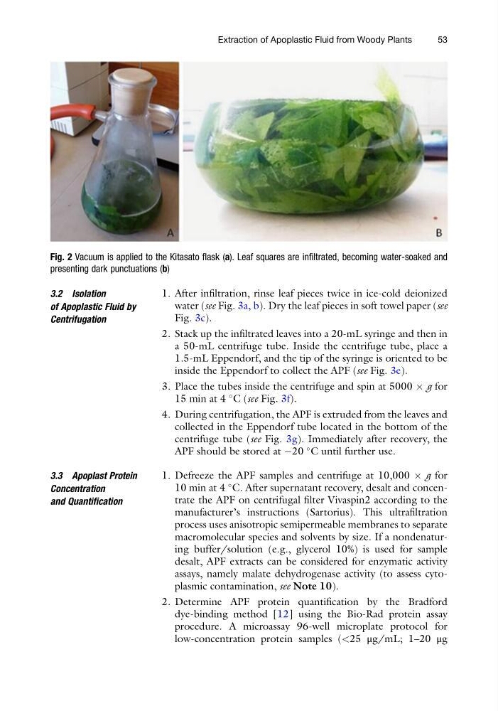
Fig. 2 Vacuum is applied to the Kitasato flask (a). Leaf squares are infiltrated, becoming water-soaked and
presenting dark punctuations (b)
Extraction of Apoplastic Fluid from Woody Plants
53
of Apoplastic Fluid by
Centrifugation
1. After infiltration, rinse leaf pieces twice in ice-cold deionized
water (see Fig. 3a, b). Dry the leaf pieces in soft towel paper (see
Fig. 3c).
2. Stack up the infiltrated leaves into a 20-mL syringe and then in
a 50-mL centrifuge tube. Inside the centrifuge tube, place a
1.5-mL Eppendorf, and the tip of the syringe is oriented to be
inside the Eppendorf to collect the APF (see Fig. 3e).
3. Place the tubes inside the centrifuge and spin at 5000 Â g for
15 min at 4
C (see Fig. 3f).
4. During centrifugation, the APF is extruded from the leaves and
collected in the Eppendorf tube located in the bottom of the
centrifuge tube (see Fig. 3g). Immediately after recovery, the
APF should be stored at À20
C until further use.
3.3 Apoplast Protein
Concentration
and Quantification
1. Defreeze the APF samples and centrifuge at 10,000 Â g for
10 min at 4
C. After supernatant recovery, desalt and concentrate the APF on centrifugal filter Vivaspin2 according to the
manufacturer’s instructions (Sartorius). This ultrafiltration
process uses anisotropic semipermeable membranes to separate
macromolecular species and solvents by size. If a nondenaturing buffer/solution (e.g., glycerol 10%) is used for sample
desalt, APF extracts can be considered for enzymatic activity
assays, namely malate dehydrogenase activity (to assess cytoplasmic contamination, see Note 10).
2. Determine APF protein quantification by the Bradford
dye-binding method [12] using the Bio-Rad protein assay
procedure. A microassay 96-well microplate protocol for
low-concentration protein samples (<25 μg/mL; 1–20 μg
Fig. 2 Vacuum is applied to the Kitasato flask (a). Leaf squares are infiltrated, becoming water-soaked and
presenting dark punctuations (b)
Extraction of Apoplastic Fluid from Woody Plants
53