

3. The MMI cell tools software also allows the manipulation of
the laser parameters (speed, focus, and power) to get a precise
cut of the desired protrusions. The settings for LCM have to be
adjusted for each sample with the objective required to cut the
cellular protrusions of interest. Different objectives may be
used depending on the cellular protrusions of interest (see
Fig. 3).
4. The laser speed should be slowed down to avoid detaching the
cells or disturbing the cellular protrusions during the process of
cutting. This will need to be determined for each sample
depending on the types of isolated cellular protrusions. For
example, for growth cones or filopodia, which are strongly
attached to the substratum, the cut velocity can be higher
(e.g., 35 μm/s). On the other hand, for more fragile structures
like TNTs, the cut velocity should be reduced (e.g., 10 μm/s).
5. Laser focus is a way to adjust the position of the laser beam in
the Z-direction within the sample, determining the thickness of
the cut. It should neither be too low to detach the membrane
as it is cutting, nor too high to not cut the membrane at all.
This will be determined by calibrating the laser focus according
to the plane tilt of the dish. Thus, the laser focus should be
adjusted for each experiment as it may vary from dish to dish
and sample to sample.
6. The power needed to cut the sample is proportional to the
sample thickness. It is represented in the percentage of UV
light transmitted by the laser. The laser power should be set
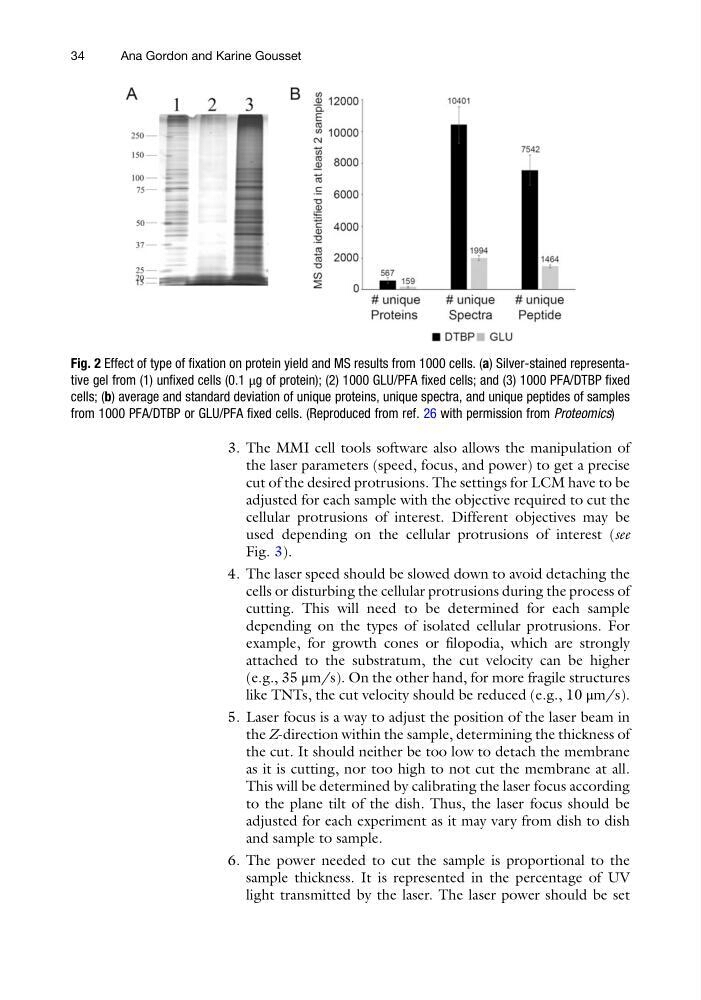
Fig. 2 Effect of type of fixation on protein yield and MS results from 1000 cells. (a) Silver-stained representative gel from (1) unfixed cells (0.1 μg of protein); (2) 1000 GLU/PFA fixed cells; and (3) 1000 PFA/DTBP fixed
cells; (b) average and standard deviation of unique proteins, unique spectra, and unique peptides of samples
from 1000 PFA/DTBP or GLU/PFA fixed cells. (Reproduced from ref. 26 with permission from Proteomics)
34
Ana Gordon and Karine Gousset
the laser parameters (speed, focus, and power) to get a precise
cut of the desired protrusions. The settings for LCM have to be
adjusted for each sample with the objective required to cut the
cellular protrusions of interest. Different objectives may be
used depending on the cellular protrusions of interest (see
Fig. 3).
4. The laser speed should be slowed down to avoid detaching the
cells or disturbing the cellular protrusions during the process of
cutting. This will need to be determined for each sample
depending on the types of isolated cellular protrusions. For
example, for growth cones or filopodia, which are strongly
attached to the substratum, the cut velocity can be higher
(e.g., 35 μm/s). On the other hand, for more fragile structures
like TNTs, the cut velocity should be reduced (e.g., 10 μm/s).
5. Laser focus is a way to adjust the position of the laser beam in
the Z-direction within the sample, determining the thickness of
the cut. It should neither be too low to detach the membrane
as it is cutting, nor too high to not cut the membrane at all.
This will be determined by calibrating the laser focus according
to the plane tilt of the dish. Thus, the laser focus should be
adjusted for each experiment as it may vary from dish to dish
and sample to sample.
6. The power needed to cut the sample is proportional to the
sample thickness. It is represented in the percentage of UV
light transmitted by the laser. The laser power should be set
Fig. 2 Effect of type of fixation on protein yield and MS results from 1000 cells. (a) Silver-stained representative gel from (1) unfixed cells (0.1 μg of protein); (2) 1000 GLU/PFA fixed cells; and (3) 1000 PFA/DTBP fixed
cells; (b) average and standard deviation of unique proteins, unique spectra, and unique peptides of samples
from 1000 PFA/DTBP or GLU/PFA fixed cells. (Reproduced from ref. 26 with permission from Proteomics)
34
Ana Gordon and Karine Gousset