

3. Incubate the samples in a thermoblock at 37
C with shaking at
1400 rpm for 5 min and sonicate for 5 min. Repeat until
complete dissolution of the protein pellet occurs. Spin.
4. Incubate with 5 μL of 200 mM TCEP in 0.2 TEAB in the
thermoblock for 1 h at 55
C.
5. Add 5 μL of 375 mM IAA to each sample and vortex them.
Incubate them at room temperature in the dark for 30 min.
6. Add 10 μL of trypsin diluted in 0.1 M TEAB to each sample in
a 1:50 (trypsin:total protein) ratio.
7. Vortex and spin and incubate overnight (not more than 16 h)
in the thermoblock at 30
C with gentle shaking at 400 rpm.
8. Prepare the TMT10plex Mass Tag Labeling Kit. Let temper at
RT for 15 min and briefly spin all the vials.
9. Add 260 μL of AcN to each vial from the commercial
TMT10plex Mass Tag Labeling Kit and vortex for 30 s.
10. Incubate for 5 min at room temperature, vortex, and spin.
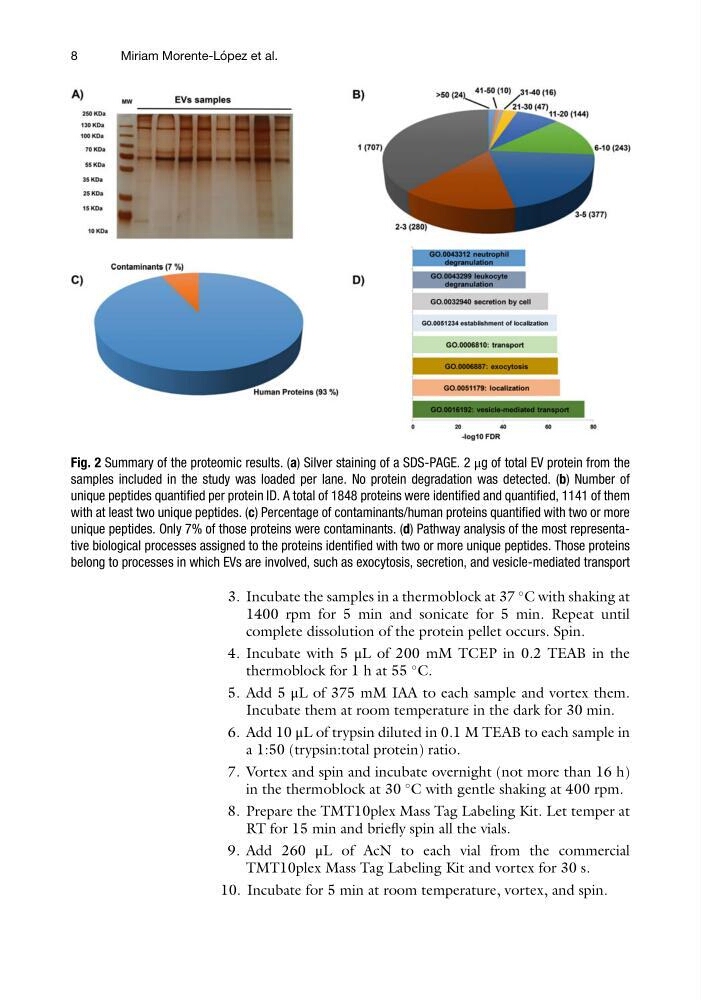
Fig. 2 Summary of the proteomic results. (a) Silver staining of a SDS-PAGE. 2 μg of total EV protein from the
samples included in the study was loaded per lane. No protein degradation was detected. (b) Number of
unique peptides quantified per protein ID. A total of 1848 proteins were identified and quantified, 1141 of them
with at least two unique peptides. (c) Percentage of contaminants/human proteins quantified with two or more
unique peptides. Only 7% of those proteins were contaminants. (d) Pathway analysis of the most representative biological processes assigned to the proteins identified with two or more unique peptides. Those proteins
belong to processes in which EVs are involved, such as exocytosis, secretion, and vesicle-mediated transport
8
Miriam Morente-Lo ´ pez et al.
C with shaking at
1400 rpm for 5 min and sonicate for 5 min. Repeat until
complete dissolution of the protein pellet occurs. Spin.
4. Incubate with 5 μL of 200 mM TCEP in 0.2 TEAB in the
thermoblock for 1 h at 55
C.
5. Add 5 μL of 375 mM IAA to each sample and vortex them.
Incubate them at room temperature in the dark for 30 min.
6. Add 10 μL of trypsin diluted in 0.1 M TEAB to each sample in
a 1:50 (trypsin:total protein) ratio.
7. Vortex and spin and incubate overnight (not more than 16 h)
in the thermoblock at 30
C with gentle shaking at 400 rpm.
8. Prepare the TMT10plex Mass Tag Labeling Kit. Let temper at
RT for 15 min and briefly spin all the vials.
9. Add 260 μL of AcN to each vial from the commercial
TMT10plex Mass Tag Labeling Kit and vortex for 30 s.
10. Incubate for 5 min at room temperature, vortex, and spin.
Fig. 2 Summary of the proteomic results. (a) Silver staining of a SDS-PAGE. 2 μg of total EV protein from the
samples included in the study was loaded per lane. No protein degradation was detected. (b) Number of
unique peptides quantified per protein ID. A total of 1848 proteins were identified and quantified, 1141 of them
with at least two unique peptides. (c) Percentage of contaminants/human proteins quantified with two or more
unique peptides. Only 7% of those proteins were contaminants. (d) Pathway analysis of the most representative biological processes assigned to the proteins identified with two or more unique peptides. Those proteins
belong to processes in which EVs are involved, such as exocytosis, secretion, and vesicle-mediated transport
8
Miriam Morente-Lo ´ pez et al.