

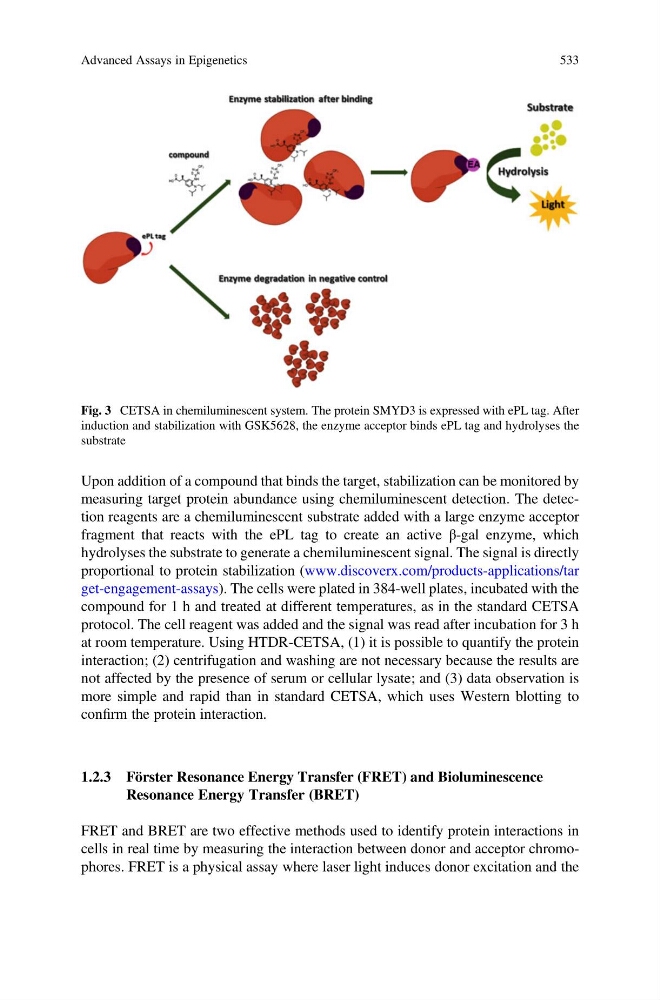
Upon addition of a compound that binds the target, stabilization can be monitored by
measuring target protein abundance using chemiluminescent detection. The detection reagents are a chemiluminescent substrate added with a large enzyme acceptor
fragment that reacts with the ePL tag to create an active β-gal enzyme, which
hydrolyses the substrate to generate a chemiluminescent signal. The signal is directly
proportional to protein stabilization (www.discoverx.com/products-applications/tar
get-engagement-assays). The cells were plated in 384-well plates, incubated with the
compound for 1 h and treated at different temperatures, as in the standard CETSA
protocol. The cell reagent was added and the signal was read after incubation for 3 h
at room temperature. Using HTDR-CETSA, (1) it is possible to quantify the protein
interaction; (2) centrifugation and washing are not necessary because the results are
not affected by the presence of serum or cellular lysate; and (3) data observation is
more simple and rapid than in standard CETSA, which uses Western blotting to
confirm the protein interaction.
1.2.3 Förster Resonance Energy Transfer (FRET) and Bioluminescence
Resonance Energy Transfer (BRET)
FRET and BRET are two effective methods used to identify protein interactions in
cells in real time by measuring the interaction between donor and acceptor chromophores. FRET is a physical assay where laser light induces donor excitation and the
Fig. 3 CETSA in chemiluminescent system. The protein SMYD3 is expressed with ePL tag. After
induction and stabilization with GSK5628, the enzyme acceptor binds ePL tag and hydrolyses the
substrate
Advanced Assays in Epigenetics
533
measuring target protein abundance using chemiluminescent detection. The detection reagents are a chemiluminescent substrate added with a large enzyme acceptor
fragment that reacts with the ePL tag to create an active β-gal enzyme, which
hydrolyses the substrate to generate a chemiluminescent signal. The signal is directly
proportional to protein stabilization (www.discoverx.com/products-applications/tar
get-engagement-assays). The cells were plated in 384-well plates, incubated with the
compound for 1 h and treated at different temperatures, as in the standard CETSA
protocol. The cell reagent was added and the signal was read after incubation for 3 h
at room temperature. Using HTDR-CETSA, (1) it is possible to quantify the protein
interaction; (2) centrifugation and washing are not necessary because the results are
not affected by the presence of serum or cellular lysate; and (3) data observation is
more simple and rapid than in standard CETSA, which uses Western blotting to
confirm the protein interaction.
1.2.3 Förster Resonance Energy Transfer (FRET) and Bioluminescence
Resonance Energy Transfer (BRET)
FRET and BRET are two effective methods used to identify protein interactions in
cells in real time by measuring the interaction between donor and acceptor chromophores. FRET is a physical assay where laser light induces donor excitation and the
Fig. 3 CETSA in chemiluminescent system. The protein SMYD3 is expressed with ePL tag. After
induction and stabilization with GSK5628, the enzyme acceptor binds ePL tag and hydrolyses the
substrate
Advanced Assays in Epigenetics
533