

modifications that are primarily used for distinguishing host DNA from foreign
pathogenic DNA [25, 26]. First indirect evidence for the presence of N6MedA in
mammals dates to 1983 [27]. Although subsequent studies by Ratel et al. were unable to
confirm the presence of N6MedA in mammals [28], more recent reports confirmed the
presence of N6MedA in mouse and human genomes [26, 29] and identified the
methylase that creates N6MedA, N6AMT1 [30]. N6MedA was shown to be widely
distributed across the genome, but depleted in exonic regions [26]. Furthermore,
N6MedA is preferentially found at TAGG sites and has been implicated in gene
repression [26, 29]. Many features of N6MedA in mammals remain to be investigated,
including the identities of possible N6MedA-interacting proteins [26].
N
4 -methyl-2
0 -deoxycytidine (4mC, Fig. 1) was first detected in the DNA of
thermophilic bacteria and bacterial mesophiles [31]. 4mC is generated by the N-4
cytosine-specific DNA methyltransferase which specifically methylates the amino
group at the N-4 position of cytosine [32]. 4mC is a part of a bacterial restrictionmodification system used to defend against foreign DNA, with several 4mC
methyltransferases and 4mC-sensitive restriction endonucleases identified in various
bacterial strains [33]. However, additional biological functions of 4mC have not
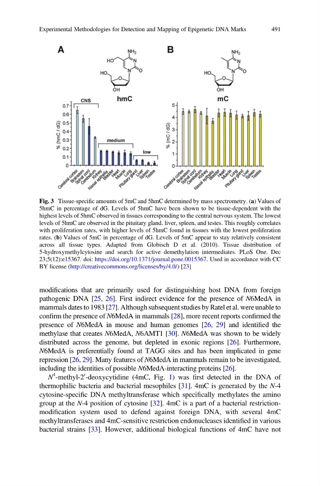
Fig. 3 Tissue-specific amounts of 5mC and 5hmC determined by mass spectrometry. (a) Values of
5hmC in percentage of dG. Levels of 5hmC have been shown to be tissue-dependent with the
highest levels of 5hmC observed in tissues corresponding to the central nervous system. The lowest
levels of 5hmC are observed in the pituitary gland, liver, spleen, and testes. This roughly correlates
with proliferation rates, with higher levels of 5hmC found in tissues with the lowest proliferation
rates. (b) Values of 5mC in percentage of dG. Levels of 5mC appear to stay relatively consistent
across all tissue types. Adapted from Globisch D et al. (2010). Tissue distribution of
5-hydroxymethylcytosine and search for active demethylation intermediates. PLoS One. Dec
23;5(12):e15367. doi: https://doi.org/10.1371/journal.pone.0015367. Used in accordance with CC
BY license (http://creativecommons.org/licenses/by/4.0/) [23]
Experimental Methodologies for Detection and Mapping of Epigenetic DNA Marks
491
pathogenic DNA [25, 26]. First indirect evidence for the presence of N6MedA in
mammals dates to 1983 [27]. Although subsequent studies by Ratel et al. were unable to
confirm the presence of N6MedA in mammals [28], more recent reports confirmed the
presence of N6MedA in mouse and human genomes [26, 29] and identified the
methylase that creates N6MedA, N6AMT1 [30]. N6MedA was shown to be widely
distributed across the genome, but depleted in exonic regions [26]. Furthermore,
N6MedA is preferentially found at TAGG sites and has been implicated in gene
repression [26, 29]. Many features of N6MedA in mammals remain to be investigated,
including the identities of possible N6MedA-interacting proteins [26].
N
4 -methyl-2
0 -deoxycytidine (4mC, Fig. 1) was first detected in the DNA of
thermophilic bacteria and bacterial mesophiles [31]. 4mC is generated by the N-4
cytosine-specific DNA methyltransferase which specifically methylates the amino
group at the N-4 position of cytosine [32]. 4mC is a part of a bacterial restrictionmodification system used to defend against foreign DNA, with several 4mC
methyltransferases and 4mC-sensitive restriction endonucleases identified in various
bacterial strains [33]. However, additional biological functions of 4mC have not
Fig. 3 Tissue-specific amounts of 5mC and 5hmC determined by mass spectrometry. (a) Values of
5hmC in percentage of dG. Levels of 5hmC have been shown to be tissue-dependent with the
highest levels of 5hmC observed in tissues corresponding to the central nervous system. The lowest
levels of 5hmC are observed in the pituitary gland, liver, spleen, and testes. This roughly correlates
with proliferation rates, with higher levels of 5hmC found in tissues with the lowest proliferation
rates. (b) Values of 5mC in percentage of dG. Levels of 5mC appear to stay relatively consistent
across all tissue types. Adapted from Globisch D et al. (2010). Tissue distribution of
5-hydroxymethylcytosine and search for active demethylation intermediates. PLoS One. Dec
23;5(12):e15367. doi: https://doi.org/10.1371/journal.pone.0015367. Used in accordance with CC
BY license (http://creativecommons.org/licenses/by/4.0/) [23]
Experimental Methodologies for Detection and Mapping of Epigenetic DNA Marks
491