

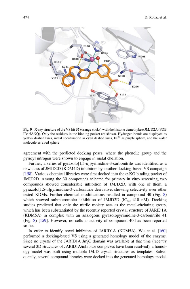
agreement with the predicted docking poses, where the phenolic group and the
pyridyl nitrogen were shown to engage in metal chelation.
Further, a series of pyrazolo[1,5-a]pyrimidine-3-carbonitrile was identified as a
new class of JMJD2D (KDM4D) inhibitors by another docking-based VS campaign
[158]. Various chemical libraries were first docked into the α-KG binding pocket of
JMJD2D. Among the 30 compounds selected for primary in vitro screening, two
compounds showed considerable inhibition of JMJD2D, with one of them, a
pyrazolo[1,5-a]pyrimidine-3-carbonitrile derivative, showing selectivity over other
tested KDMs. Further chemical modifications resulted in compound 40 (Fig. 8)
which showed submicromolar inhibition of JMJD2D (IC 50 410 nM). Docking
studies predicted that only the nitrile moiety acts as the metal-chelating group,
which has been substantiated by the recently reported crystal structure of JARID1A
(KDM5A) in complex with an analogous pyrazolopyrimidine-3-carbonitrile 41
(Fig. 8) [159]. However, no cellular activity of compound 40 has been reported
so far.
In order to identify novel inhibitors of JARID1A (KDM5A), Wu et al. [160]
performed a docking-based VS using a generated homology model of the enzyme.
Since no crystal of the JARID1A JmjC domain was available at that time (recently
several 3D structures of JARID1A/inhibitor complexes have been resolved), a homology model was built using multiple JMJD crystal structures as templates. Subsequently, several compound libraries were docked into the generated homology model.
Fig. 9 X-ray structure of the VS hit 37 (orange sticks) with the histone demethylase JMJD2A (PDB
ID: 5ANQ). Only the residues in the binding pocket are shown. Hydrogen bonds are displayed as
yellow dashed lines, metal coordination as cyan dashed lines, Fe
2+ as purple sphere, and the water
molecule as a red sphere
474
D. Robaa et al.
pyridyl nitrogen were shown to engage in metal chelation.
Further, a series of pyrazolo[1,5-a]pyrimidine-3-carbonitrile was identified as a
new class of JMJD2D (KDM4D) inhibitors by another docking-based VS campaign
[158]. Various chemical libraries were first docked into the α-KG binding pocket of
JMJD2D. Among the 30 compounds selected for primary in vitro screening, two
compounds showed considerable inhibition of JMJD2D, with one of them, a
pyrazolo[1,5-a]pyrimidine-3-carbonitrile derivative, showing selectivity over other
tested KDMs. Further chemical modifications resulted in compound 40 (Fig. 8)
which showed submicromolar inhibition of JMJD2D (IC 50 410 nM). Docking
studies predicted that only the nitrile moiety acts as the metal-chelating group,
which has been substantiated by the recently reported crystal structure of JARID1A
(KDM5A) in complex with an analogous pyrazolopyrimidine-3-carbonitrile 41
(Fig. 8) [159]. However, no cellular activity of compound 40 has been reported
so far.
In order to identify novel inhibitors of JARID1A (KDM5A), Wu et al. [160]
performed a docking-based VS using a generated homology model of the enzyme.
Since no crystal of the JARID1A JmjC domain was available at that time (recently
several 3D structures of JARID1A/inhibitor complexes have been resolved), a homology model was built using multiple JMJD crystal structures as templates. Subsequently, several compound libraries were docked into the generated homology model.
Fig. 9 X-ray structure of the VS hit 37 (orange sticks) with the histone demethylase JMJD2A (PDB
ID: 5ANQ). Only the residues in the binding pocket are shown. Hydrogen bonds are displayed as
yellow dashed lines, metal coordination as cyan dashed lines, Fe
2+ as purple sphere, and the water
molecule as a red sphere
474
D. Robaa et al.