

An iterative VS campaign combined with rational structural optimization led to
the discovery of another potent class of JMJD2A (KDM4A) inhibitors [155]. Several
compound databases were first filtered for molecules containing a metal
ion-chelating moiety. The retrieved hits were docked into the α-KG binding pocket
of JMJD2A, and the top-ranked compounds were tested in vitro against several
KDMs. This led to the identification of two 2-(pyrimidin-4-yl)pyridine-4-carboxylic
acid derivatives with promising JMJD2A inhibitory activity (IC 50 ~1 and 4 μM). A
subsequent substructure search for compounds bearing a 2-(pyrimidin-4-yl)pyridine-4-carboxylic acid scaffold resulted in a series of potent JMJD2A inhibitors
with IC 50 between 0.9 and 8.1 μM. Finally, structural optimization led to compound
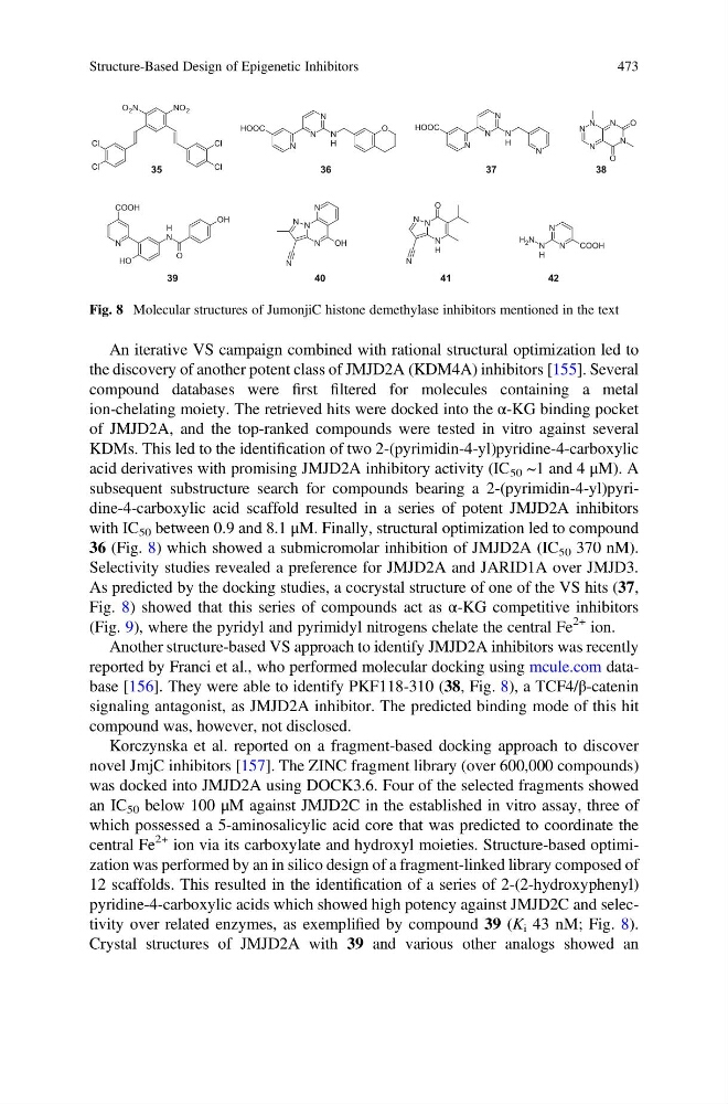
36 (Fig. 8) which showed a submicromolar inhibition of JMJD2A (IC 50 370 nM).
Selectivity studies revealed a preference for JMJD2A and JARID1A over JMJD3.
As predicted by the docking studies, a cocrystal structure of one of the VS hits (37,
Fig. 8) showed that this series of compounds act as α-KG competitive inhibitors
(Fig. 9), where the pyridyl and pyrimidyl nitrogens chelate the central Fe
2+ ion.
Another structure-based VS approach to identify JMJD2A inhibitors was recently
reported by Franci et al., who performed molecular docking using mcule.com database [156]. They were able to identify PKF118-310 (38, Fig. 8), a TCF4/β-catenin
signaling antagonist, as JMJD2A inhibitor. The predicted binding mode of this hit
compound was, however, not disclosed.
Korczynska et al. reported on a fragment-based docking approach to discover
novel JmjC inhibitors [157]. The ZINC fragment library (over 600,000 compounds)
was docked into JMJD2A using DOCK3.6. Four of the selected fragments showed
an IC 50 below 100 μM against JMJD2C in the established in vitro assay, three of
which possessed a 5-aminosalicylic acid core that was predicted to coordinate the
central Fe
2+ ion via its carboxylate and hydroxyl moieties. Structure-based optimization was performed by an in silico design of a fragment-linked library composed of
12 scaffolds. This resulted in the identification of a series of 2-(2-hydroxyphenyl)
pyridine-4-carboxylic acids which showed high potency against JMJD2C and selectivity over related enzymes, as exemplified by compound 39 (K i 43 nM; Fig. 8).
Crystal structures of JMJD2A with 39 and various other analogs showed an
35
Cl
Cl
Cl
Cl
NO 2
O 2 N
N
N
COOH
N
H
N
H 2
N
N
N
N
OH
N
N
N
N
H
N
N
HOOC
N
H
N
N
O
N
N
N
N
H
N
HOOC
O
36
37
38
39
40
N
N
N
N
N
O
O
41
N
COOH
O
H
N
H
O
OH
42
Fig. 8 Molecular structures of JumonjiC histone demethylase inhibitors mentioned in the text
Structure-Based Design of Epigenetic Inhibitors
473
the discovery of another potent class of JMJD2A (KDM4A) inhibitors [155]. Several
compound databases were first filtered for molecules containing a metal
ion-chelating moiety. The retrieved hits were docked into the α-KG binding pocket
of JMJD2A, and the top-ranked compounds were tested in vitro against several
KDMs. This led to the identification of two 2-(pyrimidin-4-yl)pyridine-4-carboxylic
acid derivatives with promising JMJD2A inhibitory activity (IC 50 ~1 and 4 μM). A
subsequent substructure search for compounds bearing a 2-(pyrimidin-4-yl)pyridine-4-carboxylic acid scaffold resulted in a series of potent JMJD2A inhibitors
with IC 50 between 0.9 and 8.1 μM. Finally, structural optimization led to compound
36 (Fig. 8) which showed a submicromolar inhibition of JMJD2A (IC 50 370 nM).
Selectivity studies revealed a preference for JMJD2A and JARID1A over JMJD3.
As predicted by the docking studies, a cocrystal structure of one of the VS hits (37,
Fig. 8) showed that this series of compounds act as α-KG competitive inhibitors
(Fig. 9), where the pyridyl and pyrimidyl nitrogens chelate the central Fe
2+ ion.
Another structure-based VS approach to identify JMJD2A inhibitors was recently
reported by Franci et al., who performed molecular docking using mcule.com database [156]. They were able to identify PKF118-310 (38, Fig. 8), a TCF4/β-catenin
signaling antagonist, as JMJD2A inhibitor. The predicted binding mode of this hit
compound was, however, not disclosed.
Korczynska et al. reported on a fragment-based docking approach to discover
novel JmjC inhibitors [157]. The ZINC fragment library (over 600,000 compounds)
was docked into JMJD2A using DOCK3.6. Four of the selected fragments showed
an IC 50 below 100 μM against JMJD2C in the established in vitro assay, three of
which possessed a 5-aminosalicylic acid core that was predicted to coordinate the
central Fe
2+ ion via its carboxylate and hydroxyl moieties. Structure-based optimization was performed by an in silico design of a fragment-linked library composed of
12 scaffolds. This resulted in the identification of a series of 2-(2-hydroxyphenyl)
pyridine-4-carboxylic acids which showed high potency against JMJD2C and selectivity over related enzymes, as exemplified by compound 39 (K i 43 nM; Fig. 8).
Crystal structures of JMJD2A with 39 and various other analogs showed an
35
Cl
Cl
Cl
Cl
NO 2
O 2 N
N
N
COOH
N
H
N
H 2
N
N
N
N
OH
N
N
N
N
H
N
N
HOOC
N
H
N
N
O
N
N
N
N
H
N
HOOC
O
36
37
38
39
40
N
N
N
N
N
O
O
41
N
COOH
O
H
N
H
O
OH
42
Fig. 8 Molecular structures of JumonjiC histone demethylase inhibitors mentioned in the text
Structure-Based Design of Epigenetic Inhibitors
473