

IC 50 values are determined with nonlinear regression fits using software such as
GraphPad or Origin. IC 50 values can be converted into K i values, which are proximal
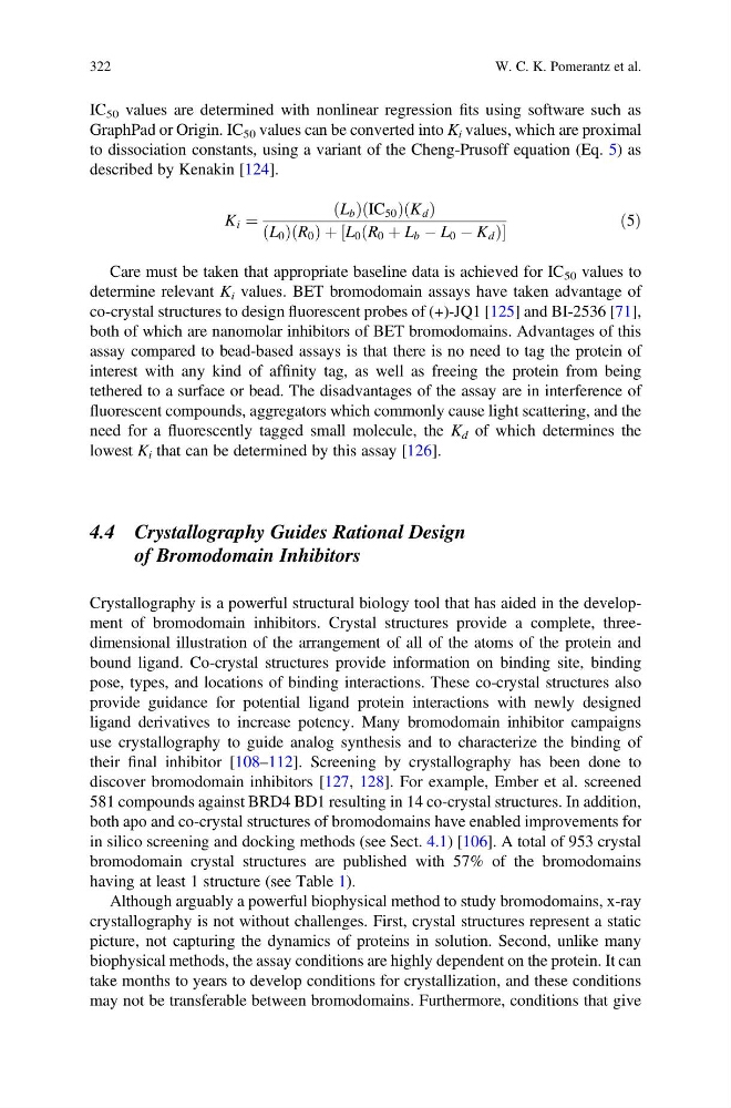
to dissociation constants, using a variant of the Cheng-Prusoff equation (Eq. 5) as
described by Kenakin [124].
K i ¼
L b
ð Þ IC 50
ð
Þ K d
ð Þ
L 0
ð Þ R 0
ð Þ þ L 0 R 0 þ L b À L 0 À K d
ð
Þ
½
ð5Þ
Care must be taken that appropriate baseline data is achieved for IC 50 values to
determine relevant K i values. BET bromodomain assays have taken advantage of
co-crystal structures to design fluorescent probes of (+)-JQ1 [125] and BI-2536 [71],
both of which are nanomolar inhibitors of BET bromodomains. Advantages of this
assay compared to bead-based assays is that there is no need to tag the protein of
interest with any kind of affinity tag, as well as freeing the protein from being
tethered to a surface or bead. The disadvantages of the assay are in interference of
fluorescent compounds, aggregators which commonly cause light scattering, and the
need for a fluorescently tagged small molecule, the K d of which determines the
lowest K i that can be determined by this assay [126].
4.4 Crystallography Guides Rational Design
of Bromodomain Inhibitors
Crystallography is a powerful structural biology tool that has aided in the development of bromodomain inhibitors. Crystal structures provide a complete, threedimensional illustration of the arrangement of all of the atoms of the protein and
bound ligand. Co-crystal structures provide information on binding site, binding
pose, types, and locations of binding interactions. These co-crystal structures also
provide guidance for potential ligand protein interactions with newly designed
ligand derivatives to increase potency. Many bromodomain inhibitor campaigns
use crystallography to guide analog synthesis and to characterize the binding of
their final inhibitor [108–112]. Screening by crystallography has been done to
discover bromodomain inhibitors [127, 128]. For example, Ember et al. screened
581 compounds against BRD4 BD1 resulting in 14 co-crystal structures. In addition,
both apo and co-crystal structures of bromodomains have enabled improvements for
in silico screening and docking methods (see Sect. 4.1) [106]. A total of 953 crystal
bromodomain crystal structures are published with 57% of the bromodomains
having at least 1 structure (see Table 1).
Although arguably a powerful biophysical method to study bromodomains, x-ray
crystallography is not without challenges. First, crystal structures represent a static
picture, not capturing the dynamics of proteins in solution. Second, unlike many
biophysical methods, the assay conditions are highly dependent on the protein. It can
take months to years to develop conditions for crystallization, and these conditions
may not be transferable between bromodomains. Furthermore, conditions that give
322
W. C. K. Pomerantz et al.
GraphPad or Origin. IC 50 values can be converted into K i values, which are proximal
to dissociation constants, using a variant of the Cheng-Prusoff equation (Eq. 5) as
described by Kenakin [124].
K i ¼
L b
ð Þ IC 50
ð
Þ K d
ð Þ
L 0
ð Þ R 0
ð Þ þ L 0 R 0 þ L b À L 0 À K d
ð
Þ
½
ð5Þ
Care must be taken that appropriate baseline data is achieved for IC 50 values to
determine relevant K i values. BET bromodomain assays have taken advantage of
co-crystal structures to design fluorescent probes of (+)-JQ1 [125] and BI-2536 [71],
both of which are nanomolar inhibitors of BET bromodomains. Advantages of this
assay compared to bead-based assays is that there is no need to tag the protein of
interest with any kind of affinity tag, as well as freeing the protein from being
tethered to a surface or bead. The disadvantages of the assay are in interference of
fluorescent compounds, aggregators which commonly cause light scattering, and the
need for a fluorescently tagged small molecule, the K d of which determines the
lowest K i that can be determined by this assay [126].
4.4 Crystallography Guides Rational Design
of Bromodomain Inhibitors
Crystallography is a powerful structural biology tool that has aided in the development of bromodomain inhibitors. Crystal structures provide a complete, threedimensional illustration of the arrangement of all of the atoms of the protein and
bound ligand. Co-crystal structures provide information on binding site, binding
pose, types, and locations of binding interactions. These co-crystal structures also
provide guidance for potential ligand protein interactions with newly designed
ligand derivatives to increase potency. Many bromodomain inhibitor campaigns
use crystallography to guide analog synthesis and to characterize the binding of
their final inhibitor [108–112]. Screening by crystallography has been done to
discover bromodomain inhibitors [127, 128]. For example, Ember et al. screened
581 compounds against BRD4 BD1 resulting in 14 co-crystal structures. In addition,
both apo and co-crystal structures of bromodomains have enabled improvements for
in silico screening and docking methods (see Sect. 4.1) [106]. A total of 953 crystal
bromodomain crystal structures are published with 57% of the bromodomains
having at least 1 structure (see Table 1).
Although arguably a powerful biophysical method to study bromodomains, x-ray
crystallography is not without challenges. First, crystal structures represent a static
picture, not capturing the dynamics of proteins in solution. Second, unlike many
biophysical methods, the assay conditions are highly dependent on the protein. It can
take months to years to develop conditions for crystallization, and these conditions
may not be transferable between bromodomains. Furthermore, conditions that give
322
W. C. K. Pomerantz et al.