

mixtures of ligands is the inability to identify which compound(s) caused the
perturbation of resonances as well as the throughput of the experiment.
A key advantage of protein-observed NMR methods is the ability to qualitatively
and quantitatively asses binding affinities. For a biomolecular binding interaction, K d
is defined as the ratio of rate constants k off /k on where k on can be estimated to be the
rate of diffusion for small molecules (although this may not always be true). Thus,
K d can be estimated from observing k off . On the NMR timescale, compounds sample
the protein-bound and unbound states at different rates that can be classified as fast,
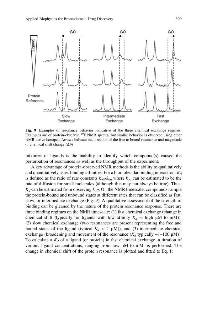
slow, or intermediate exchange (Fig. 9). A qualitative assessment of the strength of
binding can be gleaned by the nature of the protein resonance response. There are
three binding regimes on the NMR timescale: (1) fast chemical exchange (change in
chemical shift (typically for ligands with low affinity K d ¼ high μM to mM)),
(2) slow chemical exchange (two resonances are present representing the free and
bound states of the ligand (typical K d < 1 μM)), and (3) intermediate chemical
exchange (broadening and movement of the resonance (K d typically ~1–100 μM)).
To calculate a K d of a ligand (or protein) in fast chemical exchange, a titration of
various ligand concentrations, ranging from low μM to mM, is performed. The
change in chemical shift of the protein resonance is plotted and fitted to Eq. 1:
Fig. 9 Examples of resonance behavior indicative of the three chemical exchange regimes.
Examples are of protein-observed
19
F NMR spectra, but similar behavior is observed using other
NMR-active isotopes. Arrows indicate the direction of the free to bound resonance and magnitude
of chemical shift change (Δδ)
Applied Biophysics for Bromodomain Drug Discovery
309
perturbation of resonances as well as the throughput of the experiment.
A key advantage of protein-observed NMR methods is the ability to qualitatively
and quantitatively asses binding affinities. For a biomolecular binding interaction, K d
is defined as the ratio of rate constants k off /k on where k on can be estimated to be the
rate of diffusion for small molecules (although this may not always be true). Thus,
K d can be estimated from observing k off . On the NMR timescale, compounds sample
the protein-bound and unbound states at different rates that can be classified as fast,
slow, or intermediate exchange (Fig. 9). A qualitative assessment of the strength of
binding can be gleaned by the nature of the protein resonance response. There are
three binding regimes on the NMR timescale: (1) fast chemical exchange (change in
chemical shift (typically for ligands with low affinity K d ¼ high μM to mM)),
(2) slow chemical exchange (two resonances are present representing the free and
bound states of the ligand (typical K d < 1 μM)), and (3) intermediate chemical
exchange (broadening and movement of the resonance (K d typically ~1–100 μM)).
To calculate a K d of a ligand (or protein) in fast chemical exchange, a titration of
various ligand concentrations, ranging from low μM to mM, is performed. The
change in chemical shift of the protein resonance is plotted and fitted to Eq. 1:
Fig. 9 Examples of resonance behavior indicative of the three chemical exchange regimes.
Examples are of protein-observed
19
F NMR spectra, but similar behavior is observed using other
NMR-active isotopes. Arrows indicate the direction of the free to bound resonance and magnitude
of chemical shift change (Δδ)
Applied Biophysics for Bromodomain Drug Discovery
309