

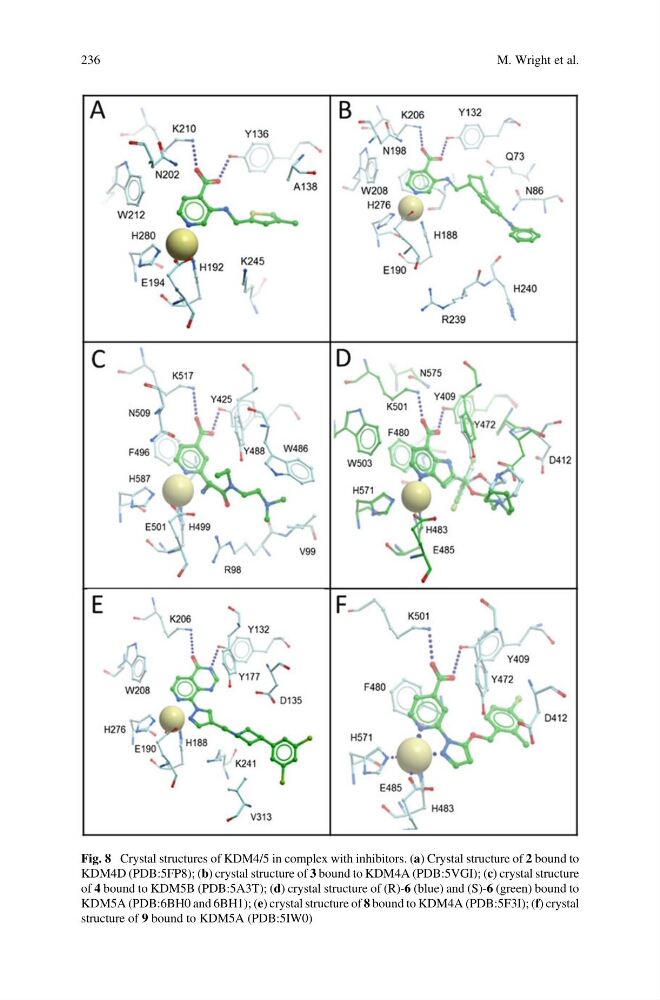
Fig. 8 Crystal structures of KDM4/5 in complex with inhibitors. (a) Crystal structure of 2 bound to
KDM4D (PDB:5FP8); (b) crystal structure of 3 bound to KDM4A (PDB:5VGI); (c) crystal structure
of 4 bound to KDM5B (PDB:5A3T); (d) crystal structure of (R)-6 (blue) and (S)-6 (green) bound to
KDM5A (PDB:6BH0 and 6BH1); (e) crystal structure of 8 bound to KDM4A (PDB:5F3I); (f) crystal
structure of 9 bound to KDM5A (PDB:5IW0)
236
M. Wright et al.
KDM4D (PDB:5FP8); (b) crystal structure of 3 bound to KDM4A (PDB:5VGI); (c) crystal structure
of 4 bound to KDM5B (PDB:5A3T); (d) crystal structure of (R)-6 (blue) and (S)-6 (green) bound to
KDM5A (PDB:6BH0 and 6BH1); (e) crystal structure of 8 bound to KDM4A (PDB:5F3I); (f) crystal
structure of 9 bound to KDM5A (PDB:5IW0)
236
M. Wright et al.