

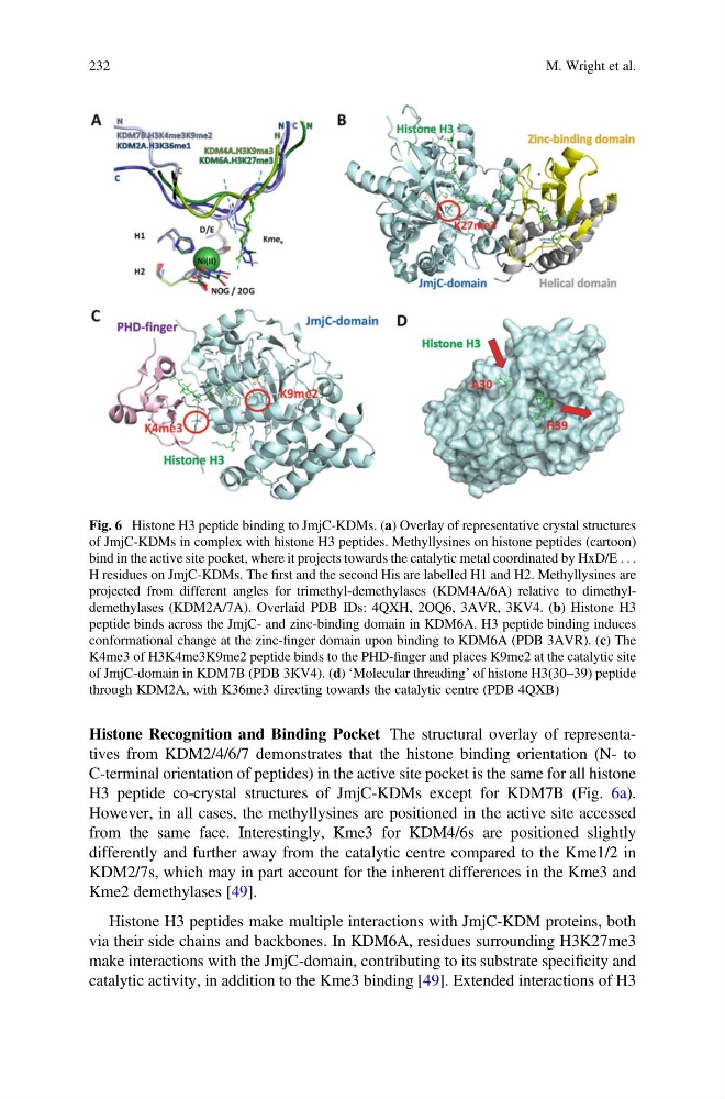
Histone Recognition and Binding Pocket The structural overlay of representatives from KDM2/4/6/7 demonstrates that the histone binding orientation (N- to
C-terminal orientation of peptides) in the active site pocket is the same for all histone
H3 peptide co-crystal structures of JmjC-KDMs except for KDM7B (Fig. 6a).
However, in all cases, the methyllysines are positioned in the active site accessed
from the same face. Interestingly, Kme3 for KDM4/6s are positioned slightly
differently and further away from the catalytic centre compared to the Kme1/2 in
KDM2/7s, which may in part account for the inherent differences in the Kme3 and
Kme2 demethylases [49].
Histone H3 peptides make multiple interactions with JmjC-KDM proteins, both
via their side chains and backbones. In KDM6A, residues surrounding H3K27me3
make interactions with the JmjC-domain, contributing to its substrate specificity and
catalytic activity, in addition to the Kme3 binding [49]. Extended interactions of H3
Fig. 6 Histone H3 peptide binding to JmjC-KDMs. (a) Overlay of representative crystal structures
of JmjC-KDMs in complex with histone H3 peptides. Methyllysines on histone peptides (cartoon)
bind in the active site pocket, where it projects towards the catalytic metal coordinated by HxD/E . . .
H residues on JmjC-KDMs. The first and the second His are labelled H1 and H2. Methyllysines are
projected from different angles for trimethyl-demethylases (KDM4A/6A) relative to dimethyldemethylases (KDM2A/7A). Overlaid PDB IDs: 4QXH, 2OQ6, 3AVR, 3KV4. (b) Histone H3
peptide binds across the JmjC- and zinc-binding domain in KDM6A. H3 peptide binding induces
conformational change at the zinc-finger domain upon binding to KDM6A (PDB 3AVR). (c) The
K4me3 of H3K4me3K9me2 peptide binds to the PHD-finger and places K9me2 at the catalytic site
of JmjC-domain in KDM7B (PDB 3KV4). (d) ‘Molecular threading’ of histone H3(30–39) peptide
through KDM2A, with K36me3 directing towards the catalytic centre (PDB 4QXB)
232
M. Wright et al.
C-terminal orientation of peptides) in the active site pocket is the same for all histone
H3 peptide co-crystal structures of JmjC-KDMs except for KDM7B (Fig. 6a).
However, in all cases, the methyllysines are positioned in the active site accessed
from the same face. Interestingly, Kme3 for KDM4/6s are positioned slightly
differently and further away from the catalytic centre compared to the Kme1/2 in
KDM2/7s, which may in part account for the inherent differences in the Kme3 and
Kme2 demethylases [49].
Histone H3 peptides make multiple interactions with JmjC-KDM proteins, both
via their side chains and backbones. In KDM6A, residues surrounding H3K27me3
make interactions with the JmjC-domain, contributing to its substrate specificity and
catalytic activity, in addition to the Kme3 binding [49]. Extended interactions of H3
Fig. 6 Histone H3 peptide binding to JmjC-KDMs. (a) Overlay of representative crystal structures
of JmjC-KDMs in complex with histone H3 peptides. Methyllysines on histone peptides (cartoon)
bind in the active site pocket, where it projects towards the catalytic metal coordinated by HxD/E . . .
H residues on JmjC-KDMs. The first and the second His are labelled H1 and H2. Methyllysines are
projected from different angles for trimethyl-demethylases (KDM4A/6A) relative to dimethyldemethylases (KDM2A/7A). Overlaid PDB IDs: 4QXH, 2OQ6, 3AVR, 3KV4. (b) Histone H3
peptide binds across the JmjC- and zinc-binding domain in KDM6A. H3 peptide binding induces
conformational change at the zinc-finger domain upon binding to KDM6A (PDB 3AVR). (c) The
K4me3 of H3K4me3K9me2 peptide binds to the PHD-finger and places K9me2 at the catalytic site
of JmjC-domain in KDM7B (PDB 3KV4). (d) ‘Molecular threading’ of histone H3(30–39) peptide
through KDM2A, with K36me3 directing towards the catalytic centre (PDB 4QXB)
232
M. Wright et al.