

KDM2A/KDM7A dimethyllysine demethylases. Thus it is proposed that this Tyr may
be important in Kme3 recognition [49].
In KDM7B, a Nε-methyl group of H3K9me2 points towards the aromatic ring of
Y234 and the other towards D249 and N333 forming hydrogen bonds. The lone pair
from the terminal di-methylated nitrogen forms a hydrogen bond with an oxygen
atom of 2OG (NOG in crystal structure); thus a steric clash prevents accommodation
of a tri-methyl lysine [8] (Fig. 4e).
In contrast, the crystal structure of mouse Kdm2A in complex with H3K36me3/
me2/me1 reveals that the spatial constraint within the methyllysine binding pocket is
not the sole determinant of methylation state selectivity in all JmjC-KDMs
[42]. Kdm2A is a H3K36me2 di-/mono-demethylase, which is structurally very
similar to KDM7s. In H3K36me2 complexed structure, the lysine side-chain binding
is stabilised by van der Waals interactions with aromatic (F215, Y199) and hydrophobic interactions (L201, I144) with dimethyl-ammonium group directed towards
D214, N298 and NOG (Fig. 4f). However, H3K36me3, which is not a substrate of
Kdm2A, can also bind in the same catalytic pocket in a similar manner. Intriguingly,
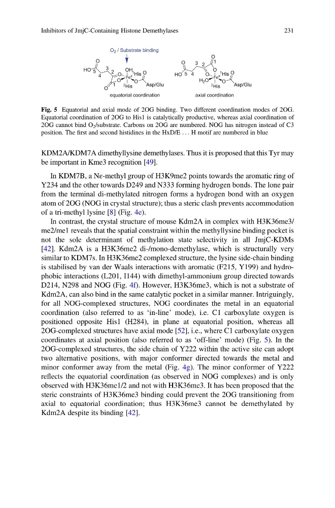
for all NOG-complexed structures, NOG coordinates the metal in an equatorial
coordination (also referred to as ‘in-line’ mode), i.e. C1 carboxylate oxygen is
positioned opposite His1 (H284), in plane at equatorial position, whereas all
2OG-complexed structures have axial mode [52], i.e., where C1 carboxylate oxygen
coordinates at axial position (also referred to as ‘off-line’ mode) (Fig. 5). In the
2OG-complexed structures, the side chain of Y222 within the active site can adopt
two alternative positions, with major conformer directed towards the metal and
minor conformer away from the metal (Fig. 4g). The minor conformer of Y222
reflects the equatorial coordination (as observed in NOG complexes) and is only
observed with H3K36me1/2 and not with H3K36me3. It has been proposed that the
steric constraints of H3K36me3 binding could prevent the 2OG transitioning from
axial to equatorial coordination; thus H3K36me3 cannot be demethylated by
Kdm2A despite its binding [42].
Fe II
O
O
1 His
O
2 His
O
Asp/Glu
O
OH
equatorial coordination
HO
1
2
3
4
O
5
Fe II
H 2 O
O
1 His
O
2 His
O
Asp/Glu
O
axial coordination
O
O
HO
1
2
3
4
5
O 2 / Substrate binding
Fig. 5 Equatorial and axial mode of 2OG binding. Two different coordination modes of 2OG.
Equatorial coordination of 2OG to His1 is catalytically productive, whereas axial coordination of
2OG cannot bind O 2 /substrate. Carbons on 2OG are numbered. NOG has nitrogen instead of C3
position. The first and second histidines in the HxD/E . . . H motif are numbered in blue
Inhibitors of JmjC-Containing Histone Demethylases
231
be important in Kme3 recognition [49].
In KDM7B, a Nε-methyl group of H3K9me2 points towards the aromatic ring of
Y234 and the other towards D249 and N333 forming hydrogen bonds. The lone pair
from the terminal di-methylated nitrogen forms a hydrogen bond with an oxygen
atom of 2OG (NOG in crystal structure); thus a steric clash prevents accommodation
of a tri-methyl lysine [8] (Fig. 4e).
In contrast, the crystal structure of mouse Kdm2A in complex with H3K36me3/
me2/me1 reveals that the spatial constraint within the methyllysine binding pocket is
not the sole determinant of methylation state selectivity in all JmjC-KDMs
[42]. Kdm2A is a H3K36me2 di-/mono-demethylase, which is structurally very
similar to KDM7s. In H3K36me2 complexed structure, the lysine side-chain binding
is stabilised by van der Waals interactions with aromatic (F215, Y199) and hydrophobic interactions (L201, I144) with dimethyl-ammonium group directed towards
D214, N298 and NOG (Fig. 4f). However, H3K36me3, which is not a substrate of
Kdm2A, can also bind in the same catalytic pocket in a similar manner. Intriguingly,
for all NOG-complexed structures, NOG coordinates the metal in an equatorial
coordination (also referred to as ‘in-line’ mode), i.e. C1 carboxylate oxygen is
positioned opposite His1 (H284), in plane at equatorial position, whereas all
2OG-complexed structures have axial mode [52], i.e., where C1 carboxylate oxygen
coordinates at axial position (also referred to as ‘off-line’ mode) (Fig. 5). In the
2OG-complexed structures, the side chain of Y222 within the active site can adopt
two alternative positions, with major conformer directed towards the metal and
minor conformer away from the metal (Fig. 4g). The minor conformer of Y222
reflects the equatorial coordination (as observed in NOG complexes) and is only
observed with H3K36me1/2 and not with H3K36me3. It has been proposed that the
steric constraints of H3K36me3 binding could prevent the 2OG transitioning from
axial to equatorial coordination; thus H3K36me3 cannot be demethylated by
Kdm2A despite its binding [42].
Fe II
O
O
1 His
O
2 His
O
Asp/Glu
O
OH
equatorial coordination
HO
1
2
3
4
O
5
Fe II
H 2 O
O
1 His
O
2 His
O
Asp/Glu
O
axial coordination
O
O
HO
1
2
3
4
5
O 2 / Substrate binding
Fig. 5 Equatorial and axial mode of 2OG binding. Two different coordination modes of 2OG.
Equatorial coordination of 2OG to His1 is catalytically productive, whereas axial coordination of
2OG cannot bind O 2 /substrate. Carbons on 2OG are numbered. NOG has nitrogen instead of C3
position. The first and second histidines in the HxD/E . . . H motif are numbered in blue
Inhibitors of JmjC-Containing Histone Demethylases
231