

5 PRMT3
5.1 Background
PRMT3 was identified by Herschman and co-workers in 1998 based on sequence
similarity with PRMT1 [11]. The crystal structure of the core of PRMT3, resolved
shortly thereafter, revealed the highly conserved active site residues and its dimeric
nature [107]. PRMT3 is a type I PRMT, predominantly present in the cytoplasm.
PRMT3 contains a zinc finger for substrate recognition with known substrates
including the 40S ribosomal protein S2, tumour protein p53 and (at least in vitro)
histone H4R3 [108]. A recent screening via bio-orthogonal profiling of protein
methylation using engineered methyltransferases resulted in the identification of
over 80 substrates of PRMT3, 70% of which were cytoplasmic [109]. However,
their functions in a biological setting remain to be elucidated. The human tumour
suppressor protein DAL-1/4.1B (differentially expressed in adenocarcinoma of the
lung) has also been shown to interact with PRMT3, inhibiting its methylating
activity and leading to apoptosis in breast cancer cells [110].
5.2 Inhibitors: In Vitro and Cell-Based Activities
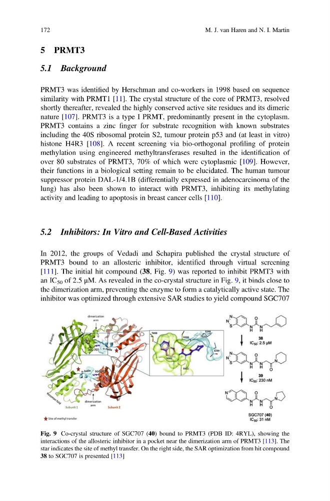
In 2012, the groups of Vedadi and Schapira published the crystal structure of
PRMT3 bound to an allosteric inhibitor, identified through virtual screening
[111]. The initial hit compound (38, Fig. 9) was reported to inhibit PRMT3 with
an IC 50 of 2.5 μM. As revealed in the co-crystal structure in Fig. 9, it binds close to
the dimerization arm, preventing the enzyme to form a catalytically active state. The
inhibitor was optimized through extensive SAR studies to yield compound SGC707
Fig. 9 Co-crystal structure of SGC707 (40) bound to PRMT3 (PDB ID: 4RYL), showing the
interactions of the allosteric inhibitor in a pocket near the dimerization arm of PRMT3 [113]. The
star indicates the site of methyl transfer. On the right side, the SAR optimization from hit compound
38 to SGC707 is presented [113]
172
M. J. van Haren and N. I. Martin
5.1 Background
PRMT3 was identified by Herschman and co-workers in 1998 based on sequence
similarity with PRMT1 [11]. The crystal structure of the core of PRMT3, resolved
shortly thereafter, revealed the highly conserved active site residues and its dimeric
nature [107]. PRMT3 is a type I PRMT, predominantly present in the cytoplasm.
PRMT3 contains a zinc finger for substrate recognition with known substrates
including the 40S ribosomal protein S2, tumour protein p53 and (at least in vitro)
histone H4R3 [108]. A recent screening via bio-orthogonal profiling of protein
methylation using engineered methyltransferases resulted in the identification of
over 80 substrates of PRMT3, 70% of which were cytoplasmic [109]. However,
their functions in a biological setting remain to be elucidated. The human tumour
suppressor protein DAL-1/4.1B (differentially expressed in adenocarcinoma of the
lung) has also been shown to interact with PRMT3, inhibiting its methylating
activity and leading to apoptosis in breast cancer cells [110].
5.2 Inhibitors: In Vitro and Cell-Based Activities
In 2012, the groups of Vedadi and Schapira published the crystal structure of
PRMT3 bound to an allosteric inhibitor, identified through virtual screening
[111]. The initial hit compound (38, Fig. 9) was reported to inhibit PRMT3 with
an IC 50 of 2.5 μM. As revealed in the co-crystal structure in Fig. 9, it binds close to
the dimerization arm, preventing the enzyme to form a catalytically active state. The
inhibitor was optimized through extensive SAR studies to yield compound SGC707
Fig. 9 Co-crystal structure of SGC707 (40) bound to PRMT3 (PDB ID: 4RYL), showing the
interactions of the allosteric inhibitor in a pocket near the dimerization arm of PRMT3 [113]. The
star indicates the site of methyl transfer. On the right side, the SAR optimization from hit compound
38 to SGC707 is presented [113]
172
M. J. van Haren and N. I. Martin