

defined voltage. As the result of the acceleration the electrons exhibit an accurate
energy, normally 70 eV. The accelerated electrons collide with the organic molecules within the ion source.
Due to the high electron energies the collision provokes the emission of a
secondary electron from the molecules forming positive ions. The collision transfers
simultaneously additional energy towards the molecules that cannot be easily
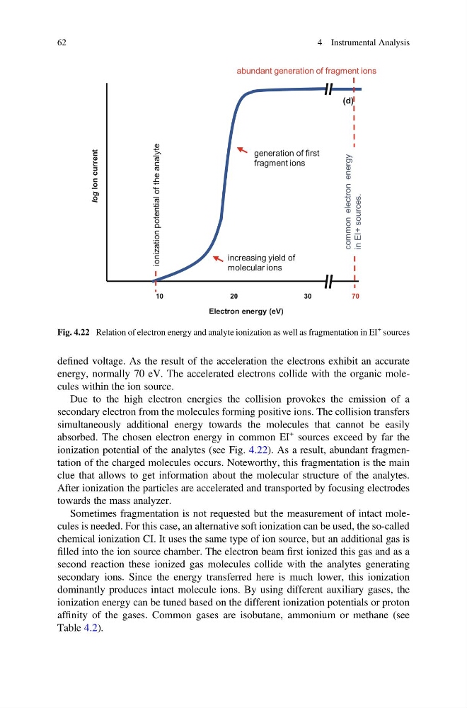
absorbed. The chosen electron energy in common EI
+ sources exceed by far the
ionization potential of the analytes (see Fig. 4.22). As a result, abundant fragmentation of the charged molecules occurs. Noteworthy, this fragmentation is the main
clue that allows to get information about the molecular structure of the analytes.
After ionization the particles are accelerated and transported by focusing electrodes
towards the mass analyzer.
Sometimes fragmentation is not requested but the measurement of intact molecules is needed. For this case, an alternative soft ionization can be used, the so-called
chemical ionization CI. It uses the same type of ion source, but an additional gas is
filled into the ion source chamber. The electron beam first ionized this gas and as a
second reaction these ionized gas molecules collide with the analytes generating
secondary ions. Since the energy transferred here is much lower, this ionization
dominantly produces intact molecule ions. By using different auxiliary gases, the
ionization energy can be tuned based on the different ionization potentials or proton
affinity of the gases. Common gases are isobutane, ammonium or methane (see
Table 4.2).
Electron energy (eV)
10
20
30
70
(d)
log Ion current
ionization potential of the analyte
increasing yield of
molecular ions
generation of first
fragment ions
abundant generation of fragment ions
common electron energy
in EI+ sources.
Fig. 4.22 Relation of electron energy and analyte ionization as well as fragmentation in EI
+ sources
62
4 Instrumental Analysis
energy, normally 70 eV. The accelerated electrons collide with the organic molecules within the ion source.
Due to the high electron energies the collision provokes the emission of a
secondary electron from the molecules forming positive ions. The collision transfers
simultaneously additional energy towards the molecules that cannot be easily
absorbed. The chosen electron energy in common EI
+ sources exceed by far the
ionization potential of the analytes (see Fig. 4.22). As a result, abundant fragmentation of the charged molecules occurs. Noteworthy, this fragmentation is the main
clue that allows to get information about the molecular structure of the analytes.
After ionization the particles are accelerated and transported by focusing electrodes
towards the mass analyzer.
Sometimes fragmentation is not requested but the measurement of intact molecules is needed. For this case, an alternative soft ionization can be used, the so-called
chemical ionization CI. It uses the same type of ion source, but an additional gas is
filled into the ion source chamber. The electron beam first ionized this gas and as a
second reaction these ionized gas molecules collide with the analytes generating
secondary ions. Since the energy transferred here is much lower, this ionization
dominantly produces intact molecule ions. By using different auxiliary gases, the
ionization energy can be tuned based on the different ionization potentials or proton
affinity of the gases. Common gases are isobutane, ammonium or methane (see
Table 4.2).
Electron energy (eV)
10
20
30
70
(d)
log Ion current
ionization potential of the analyte
increasing yield of
molecular ions
generation of first
fragment ions
abundant generation of fragment ions
common electron energy
in EI+ sources.
Fig. 4.22 Relation of electron energy and analyte ionization as well as fragmentation in EI
+ sources
62
4 Instrumental Analysis