

3 Aptamer-Based Point of Care Testing Schemes
57
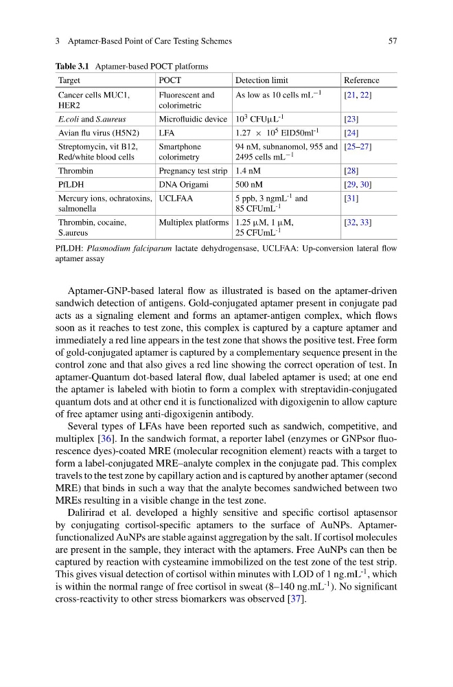
Table 3.1 Aptamer-based POCT platforms
Target
POCT
Detection limit
Reference
Cancer cells MUC1,
HER2
Fluorescent and
colorimetric
As low as 10 cells mL −1
[21, 22]
E.coli and S.aureus
Microfluidic device 10 3 CFUμL -1
[23]
Avian flu virus (H5N2)
LFA
1.27 × 10 5 EID50ml -1
[24]
Streptomycin, vit B12,
Red/white blood cells
Smartphone
colorimetry
94 nM, subnanomol, 955 and
2495 cells mL −1
[25–27]
Thrombin
Pregnancy test strip 1.4 nM
[28]
PfLDH
DNA Origami
500 nM
[29, 30]
Mercury ions, ochratoxins,
salmonella
UCLFAA
5 ppb, 3 ngmL -1 and
85 CFUmL -1
[31]
Thrombin, cocaine,
S.aureus
Multiplex platforms 1.25 μM, 1 μM,
25 CFUmL -1
[32, 33]
PfLDH: Plasmodium falciparum lactate dehydrogensase, UCLFAA: Up-conversion lateral flow
aptamer assay
Aptamer-GNP-based lateral flow as illustrated is based on the aptamer-driven
sandwich detection of antigens. Gold-conjugated aptamer present in conjugate pad
acts as a signaling element and forms an aptamer-antigen complex, which flows
soon as it reaches to test zone, this complex is captured by a capture aptamer and
immediately a red line appears in the test zone that shows the positive test. Free form
of gold-conjugated aptamer is captured by a complementary sequence present in the
control zone and that also gives a red line showing the correct operation of test. In
aptamer-Quantum dot-based lateral flow, dual labeled aptamer is used; at one end
the aptamer is labeled with biotin to form a complex with streptavidin-conjugated
quantum dots and at other end it is functionalized with digoxigenin to allow capture
of free aptamer using anti-digoxigenin antibody.
Several types of LFAs have been reported such as sandwich, competitive, and
multiplex [36]. In the sandwich format, a reporter label (enzymes or GNPsor fluorescence dyes)-coated MRE (molecular recognition element) reacts with a target to
form a label-conjugated MRE–analyte complex in the conjugate pad. This complex
travels to the test zone by capillary action and is captured by another aptamer (second
MRE) that binds in such a way that the analyte becomes sandwiched between two
MREs resulting in a visible change in the test zone.
Dalirirad et al. developed a highly sensitive and specific cortisol aptasensor
by conjugating cortisol-specific aptamers to the surface of AuNPs. Aptamerfunctionalized AuNPs are stable against aggregation by the salt. If cortisol molecules
are present in the sample, they interact with the aptamers. Free AuNPs can then be
captured by reaction with cysteamine immobilized on the test zone of the test strip.
This gives visual detection of cortisol within minutes with LOD of 1 ng.mL
-1 , which
is within the normal range of free cortisol in sweat (8–140 ng.mL
-1 ). No significant
cross-reactivity to other stress biomarkers was observed [37].
57
Table 3.1 Aptamer-based POCT platforms
Target
POCT
Detection limit
Reference
Cancer cells MUC1,
HER2
Fluorescent and
colorimetric
As low as 10 cells mL −1
[21, 22]
E.coli and S.aureus
Microfluidic device 10 3 CFUμL -1
[23]
Avian flu virus (H5N2)
LFA
1.27 × 10 5 EID50ml -1
[24]
Streptomycin, vit B12,
Red/white blood cells
Smartphone
colorimetry
94 nM, subnanomol, 955 and
2495 cells mL −1
[25–27]
Thrombin
Pregnancy test strip 1.4 nM
[28]
PfLDH
DNA Origami
500 nM
[29, 30]
Mercury ions, ochratoxins,
salmonella
UCLFAA
5 ppb, 3 ngmL -1 and
85 CFUmL -1
[31]
Thrombin, cocaine,
S.aureus
Multiplex platforms 1.25 μM, 1 μM,
25 CFUmL -1
[32, 33]
PfLDH: Plasmodium falciparum lactate dehydrogensase, UCLFAA: Up-conversion lateral flow
aptamer assay
Aptamer-GNP-based lateral flow as illustrated is based on the aptamer-driven
sandwich detection of antigens. Gold-conjugated aptamer present in conjugate pad
acts as a signaling element and forms an aptamer-antigen complex, which flows
soon as it reaches to test zone, this complex is captured by a capture aptamer and
immediately a red line appears in the test zone that shows the positive test. Free form
of gold-conjugated aptamer is captured by a complementary sequence present in the
control zone and that also gives a red line showing the correct operation of test. In
aptamer-Quantum dot-based lateral flow, dual labeled aptamer is used; at one end
the aptamer is labeled with biotin to form a complex with streptavidin-conjugated
quantum dots and at other end it is functionalized with digoxigenin to allow capture
of free aptamer using anti-digoxigenin antibody.
Several types of LFAs have been reported such as sandwich, competitive, and
multiplex [36]. In the sandwich format, a reporter label (enzymes or GNPsor fluorescence dyes)-coated MRE (molecular recognition element) reacts with a target to
form a label-conjugated MRE–analyte complex in the conjugate pad. This complex
travels to the test zone by capillary action and is captured by another aptamer (second
MRE) that binds in such a way that the analyte becomes sandwiched between two
MREs resulting in a visible change in the test zone.
Dalirirad et al. developed a highly sensitive and specific cortisol aptasensor
by conjugating cortisol-specific aptamers to the surface of AuNPs. Aptamerfunctionalized AuNPs are stable against aggregation by the salt. If cortisol molecules
are present in the sample, they interact with the aptamers. Free AuNPs can then be
captured by reaction with cysteamine immobilized on the test zone of the test strip.
This gives visual detection of cortisol within minutes with LOD of 1 ng.mL
-1 , which
is within the normal range of free cortisol in sweat (8–140 ng.mL
-1 ). No significant
cross-reactivity to other stress biomarkers was observed [37].