

34
D. Li et al.
2.2.1 Single Photon Emission Computed Tomography
(SPECT)
In 1997, Charlton et al. reported the first application of aptamer in molecular imaging
[18], a DNA aptamer (NX21909), which was an inhibitor of human neutrophil elastase secreted by activated neutrophils during inflammation. The elastase aptamer
(NX21909), a negative control aptamer and rat IgG as a positive control were radiolabeled with
99m Tc and injected intravenously into rats with induced inflammation
in a forelimb. The biodistribution of the aptamer was measured by scintigraphy. The
elastase aptamer showed longer lasting and higher signal than the negative control
in the interest area. Background intensity in peripheral tissues was low because the
clearance from circulation was more rapid compared with the positive control. Moreover, the elastase aptamer reached the peak target-to-background ratio of 4.3 ± 0.6
in 2 h. In contrast, IgG used clinically in inflammation imaging took a longer time
to achieve a lower ratio of 3.1 ± 0.1 in 3 h.
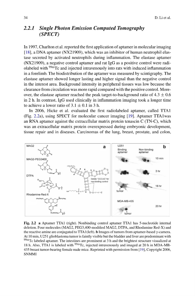
In 2006, Hicke et al. evaluated the first radiolabeled aptamer, called TTA1
(Fig. 2.2a), using SPECT for molecular cancer imaging [19]. Aptamer TTA1was
an RNA aptamer against the extracellular matrix protein tenascin C (TN-C), which
was an extracellular matrix protein overexpressed during embryonic development,
tissue repair and in diseases. Carcinomas of the lung, breast, prostate, and colon,
Fig. 2.2 a Aptamer TTA1 (right). Nonbinding control aptamer TTA1 has 5-nucleotide internal
deletion. Four molecules (MAG2, PEG3,400-modified MAG2, DTPA, and Rhodamine Red-X) and
the reactive amine are conjugated to TTA1(left). b Images of tumors from aptamer-based γ-camera.
At 10 min, U251 glioblastoma tumor is faintly visible but the bladder and liver are predominant with
99m Tc labeled aptamer. The intestines are prominent at 3 h and the brightest structure visualized at
18 h. Also, TTA1 is labeled with 99m Tc, injected intravenously and imaged at 20 h in MDA-MB435 breast tumor-bearing female nude mice. Reprinted with permission from [19], Copyright 2006,
SNMMI
D. Li et al.
2.2.1 Single Photon Emission Computed Tomography
(SPECT)
In 1997, Charlton et al. reported the first application of aptamer in molecular imaging
[18], a DNA aptamer (NX21909), which was an inhibitor of human neutrophil elastase secreted by activated neutrophils during inflammation. The elastase aptamer
(NX21909), a negative control aptamer and rat IgG as a positive control were radiolabeled with
99m Tc and injected intravenously into rats with induced inflammation
in a forelimb. The biodistribution of the aptamer was measured by scintigraphy. The
elastase aptamer showed longer lasting and higher signal than the negative control
in the interest area. Background intensity in peripheral tissues was low because the
clearance from circulation was more rapid compared with the positive control. Moreover, the elastase aptamer reached the peak target-to-background ratio of 4.3 ± 0.6
in 2 h. In contrast, IgG used clinically in inflammation imaging took a longer time
to achieve a lower ratio of 3.1 ± 0.1 in 3 h.
In 2006, Hicke et al. evaluated the first radiolabeled aptamer, called TTA1
(Fig. 2.2a), using SPECT for molecular cancer imaging [19]. Aptamer TTA1was
an RNA aptamer against the extracellular matrix protein tenascin C (TN-C), which
was an extracellular matrix protein overexpressed during embryonic development,
tissue repair and in diseases. Carcinomas of the lung, breast, prostate, and colon,
Fig. 2.2 a Aptamer TTA1 (right). Nonbinding control aptamer TTA1 has 5-nucleotide internal
deletion. Four molecules (MAG2, PEG3,400-modified MAG2, DTPA, and Rhodamine Red-X) and
the reactive amine are conjugated to TTA1(left). b Images of tumors from aptamer-based γ-camera.
At 10 min, U251 glioblastoma tumor is faintly visible but the bladder and liver are predominant with
99m Tc labeled aptamer. The intestines are prominent at 3 h and the brightest structure visualized at
18 h. Also, TTA1 is labeled with 99m Tc, injected intravenously and imaged at 20 h in MDA-MB435 breast tumor-bearing female nude mice. Reprinted with permission from [19], Copyright 2006,
SNMMI