

13 Some Frontier Technologies for Aptamers in Medical Applications
383
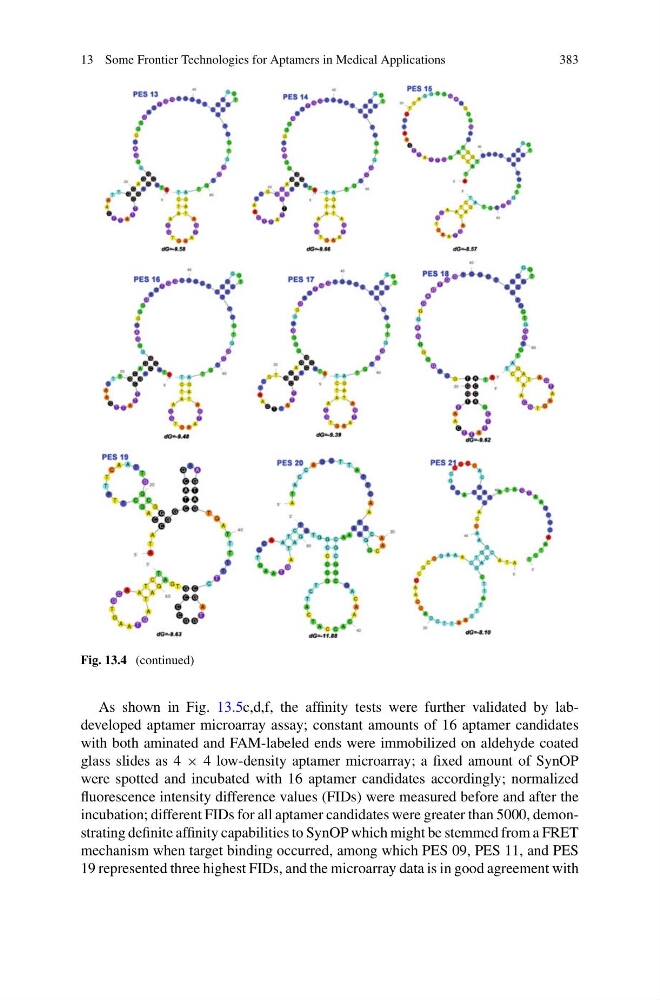
Fig. 13.4 (continued)
As shown in Fig. 13.5c,d,f, the affinity tests were further validated by labdeveloped aptamer microarray assay; constant amounts of 16 aptamer candidates
with both aminated and FAM-labeled ends were immobilized on aldehyde coated
glass slides as 4 × 4 low-density aptamer microarray; a fixed amount of SynOP
were spotted and incubated with 16 aptamer candidates accordingly; normalized
fluorescence intensity difference values (FIDs) were measured before and after the
incubation; different FIDs for all aptamer candidates were greater than 5000, demonstrating definite affinity capabilities to SynOP which might be stemmed from a FRET
mechanism when target binding occurred, among which PES 09, PES 11, and PES
19 represented three highest FIDs, and the microarray data is in good agreement with
383
Fig. 13.4 (continued)
As shown in Fig. 13.5c,d,f, the affinity tests were further validated by labdeveloped aptamer microarray assay; constant amounts of 16 aptamer candidates
with both aminated and FAM-labeled ends were immobilized on aldehyde coated
glass slides as 4 × 4 low-density aptamer microarray; a fixed amount of SynOP
were spotted and incubated with 16 aptamer candidates accordingly; normalized
fluorescence intensity difference values (FIDs) were measured before and after the
incubation; different FIDs for all aptamer candidates were greater than 5000, demonstrating definite affinity capabilities to SynOP which might be stemmed from a FRET
mechanism when target binding occurred, among which PES 09, PES 11, and PES
19 represented three highest FIDs, and the microarray data is in good agreement with