

8 Aptamers for the Diagnosis of Infectious Diseases
219
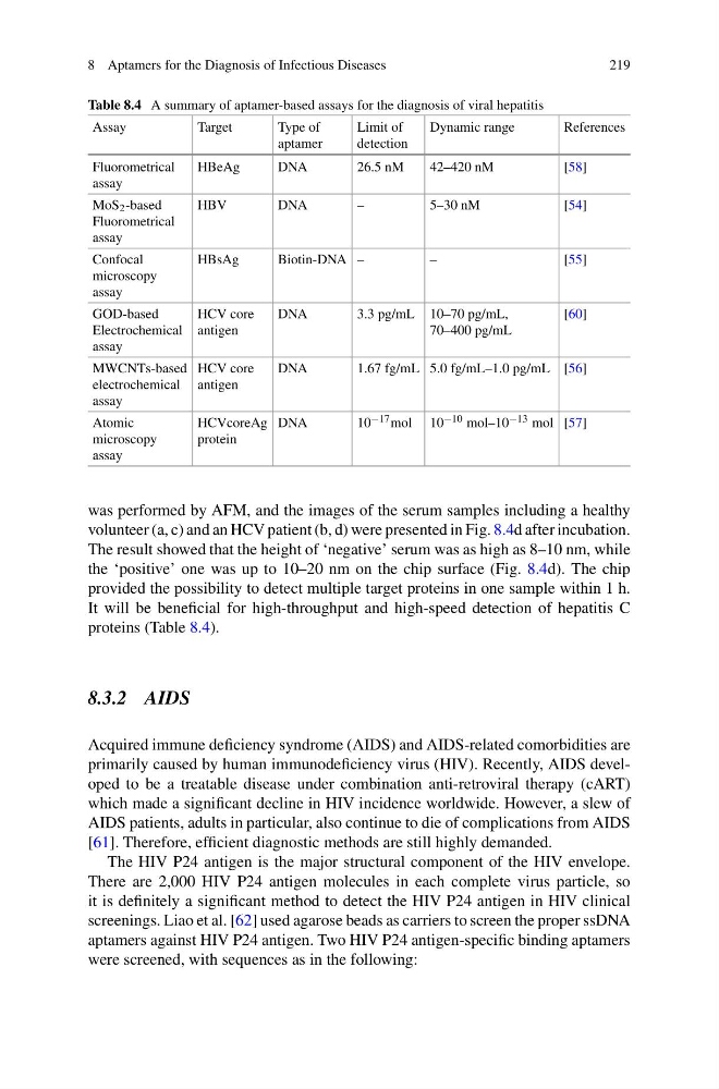
Table 8.4 A summary of aptamer-based assays for the diagnosis of viral hepatitis
Assay
Target
Type of
aptamer
Limit of
detection
Dynamic range
References
Fluorometrical
assay
HBeAg
DNA
26.5 nM
42–420 nM
[58]
MoS 2 -based
Fluorometrical
assay
HBV
DNA
–
5–30 nM
[54]
Confocal
microscopy
assay
HBsAg
Biotin-DNA –
–
[55]
GOD-based
Electrochemical
assay
HCV core
antigen
DNA
3.3 pg/mL 10–70 pg/mL,
70–400 pg/mL
[60]
MWCNTs-based
electrochemical
assay
HCV core
antigen
DNA
1.67 fg/mL 5.0 fg/mL–1.0 pg/mL [56]
Atomic
microscopy
assay
HCVcoreAg
protein
DNA
10 −17 mol 10 −10 mol–10 −13 mol [57]
was performed by AFM, and the images of the serum samples including a healthy
volunteer (a, c) and an HCV patient (b, d) were presented in Fig. 8.4d after incubation.
The result showed that the height of ‘negative’ serum was as high as 8–10 nm, while
the ‘positive’ one was up to 10–20 nm on the chip surface (Fig. 8.4d). The chip
provided the possibility to detect multiple target proteins in one sample within 1 h.
It will be beneficial for high-throughput and high-speed detection of hepatitis C
proteins (Table 8.4).
8.3.2 AIDS
Acquired immune deficiency syndrome (AIDS) and AIDS-related comorbidities are
primarily caused by human immunodeficiency virus (HIV). Recently, AIDS developed to be a treatable disease under combination anti-retroviral therapy (cART)
which made a significant decline in HIV incidence worldwide. However, a slew of
AIDS patients, adults in particular, also continue to die of complications from AIDS
[61]. Therefore, efficient diagnostic methods are still highly demanded.
The HIV P24 antigen is the major structural component of the HIV envelope.
There are 2,000 HIV P24 antigen molecules in each complete virus particle, so
it is definitely a significant method to detect the HIV P24 antigen in HIV clinical
screenings. Liao et al. [62] used agarose beads as carriers to screen the proper ssDNA
aptamers against HIV P24 antigen. Two HIV P24 antigen-specific binding aptamers
were screened, with sequences as in the following:
219
Table 8.4 A summary of aptamer-based assays for the diagnosis of viral hepatitis
Assay
Target
Type of
aptamer
Limit of
detection
Dynamic range
References
Fluorometrical
assay
HBeAg
DNA
26.5 nM
42–420 nM
[58]
MoS 2 -based
Fluorometrical
assay
HBV
DNA
–
5–30 nM
[54]
Confocal
microscopy
assay
HBsAg
Biotin-DNA –
–
[55]
GOD-based
Electrochemical
assay
HCV core
antigen
DNA
3.3 pg/mL 10–70 pg/mL,
70–400 pg/mL
[60]
MWCNTs-based
electrochemical
assay
HCV core
antigen
DNA
1.67 fg/mL 5.0 fg/mL–1.0 pg/mL [56]
Atomic
microscopy
assay
HCVcoreAg
protein
DNA
10 −17 mol 10 −10 mol–10 −13 mol [57]
was performed by AFM, and the images of the serum samples including a healthy
volunteer (a, c) and an HCV patient (b, d) were presented in Fig. 8.4d after incubation.
The result showed that the height of ‘negative’ serum was as high as 8–10 nm, while
the ‘positive’ one was up to 10–20 nm on the chip surface (Fig. 8.4d). The chip
provided the possibility to detect multiple target proteins in one sample within 1 h.
It will be beneficial for high-throughput and high-speed detection of hepatitis C
proteins (Table 8.4).
8.3.2 AIDS
Acquired immune deficiency syndrome (AIDS) and AIDS-related comorbidities are
primarily caused by human immunodeficiency virus (HIV). Recently, AIDS developed to be a treatable disease under combination anti-retroviral therapy (cART)
which made a significant decline in HIV incidence worldwide. However, a slew of
AIDS patients, adults in particular, also continue to die of complications from AIDS
[61]. Therefore, efficient diagnostic methods are still highly demanded.
The HIV P24 antigen is the major structural component of the HIV envelope.
There are 2,000 HIV P24 antigen molecules in each complete virus particle, so
it is definitely a significant method to detect the HIV P24 antigen in HIV clinical
screenings. Liao et al. [62] used agarose beads as carriers to screen the proper ssDNA
aptamers against HIV P24 antigen. Two HIV P24 antigen-specific binding aptamers
were screened, with sequences as in the following: