

218
T. Zhang et al.
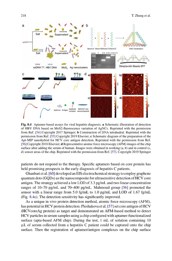
Fig. 8.4 Aptamer-based assays for viral hepatitis diagnosis. a Schematic illustration of detection
of HBV DNA based on MoS2-fluorescence variation of AgNCs. Reprinted with the permission
from Ref. [54] Copyright 2017 Springer; b Construction of DNA tetrahedral. Reprinted with the
permission from Ref. [55] Copyright 2019 Elsevier; c Schematic diagram of the preparation of the
Apt-MIP nanohybrid for HCV core antigen detection. Reprinted with the permission from Ref.
[56] Copyright 2018 Elsevier; d Representative atomic force microscopy (AFM) images of the chip
surface after adding the serum of human. Images were obtained in working (a, b) and in control (c,
d) sensor areas of the chip. Reprinted with the permission from Ref. [57]. Copyright 2019 Springer
patients do not respond to the therapy. Specific aptamers based on core protein has
held promising prospects in the early diagnosis of hepatitis C patients.
Ghanbari et al. [60] developed an EIS electrochemical strategy to employ graphene
quantum dots (GQDs) as the nanocomposite for ultrasensitive detection of HCV core
antigen. The strategy achieved a low LOD of 3.3 pg/mL and two linear concentration
ranges of 10–70 pg/mL and 70–400 pg/mL. Mahmoud group [56] promoted the
sensor with a linear range from 5.0 fg/mL to 1.0 pg/mL and LOD of 1.67 fg/mL
(Fig. 8.4c). The detection sensitivity has significantly improved.
As a unique in vivo protein detection method, atomic force microscopy (AFM),
has potential in HCV protein detection. Pleshakova et al. [57] set core antigen of HCV
(HCVcoreAg protein) as target and demonstrated an AFM-based method to detect
HCV particles in serum samples using a chip configured with aptamer-functionalized
surface (apta-based AFM chip). During the test, 1 mL of solution containing 10
µL of serum collected from a hepatitis C patient could be captured onto the chip
surface. Then the registration of aptamer/antigen complexes on the chip surface
T. Zhang et al.
Fig. 8.4 Aptamer-based assays for viral hepatitis diagnosis. a Schematic illustration of detection
of HBV DNA based on MoS2-fluorescence variation of AgNCs. Reprinted with the permission
from Ref. [54] Copyright 2017 Springer; b Construction of DNA tetrahedral. Reprinted with the
permission from Ref. [55] Copyright 2019 Elsevier; c Schematic diagram of the preparation of the
Apt-MIP nanohybrid for HCV core antigen detection. Reprinted with the permission from Ref.
[56] Copyright 2018 Elsevier; d Representative atomic force microscopy (AFM) images of the chip
surface after adding the serum of human. Images were obtained in working (a, b) and in control (c,
d) sensor areas of the chip. Reprinted with the permission from Ref. [57]. Copyright 2019 Springer
patients do not respond to the therapy. Specific aptamers based on core protein has
held promising prospects in the early diagnosis of hepatitis C patients.
Ghanbari et al. [60] developed an EIS electrochemical strategy to employ graphene
quantum dots (GQDs) as the nanocomposite for ultrasensitive detection of HCV core
antigen. The strategy achieved a low LOD of 3.3 pg/mL and two linear concentration
ranges of 10–70 pg/mL and 70–400 pg/mL. Mahmoud group [56] promoted the
sensor with a linear range from 5.0 fg/mL to 1.0 pg/mL and LOD of 1.67 fg/mL
(Fig. 8.4c). The detection sensitivity has significantly improved.
As a unique in vivo protein detection method, atomic force microscopy (AFM),
has potential in HCV protein detection. Pleshakova et al. [57] set core antigen of HCV
(HCVcoreAg protein) as target and demonstrated an AFM-based method to detect
HCV particles in serum samples using a chip configured with aptamer-functionalized
surface (apta-based AFM chip). During the test, 1 mL of solution containing 10
µL of serum collected from a hepatitis C patient could be captured onto the chip
surface. Then the registration of aptamer/antigen complexes on the chip surface