

Image-Based 2D PCD for Morphological Analysis of Tendrils-Like Structure
89
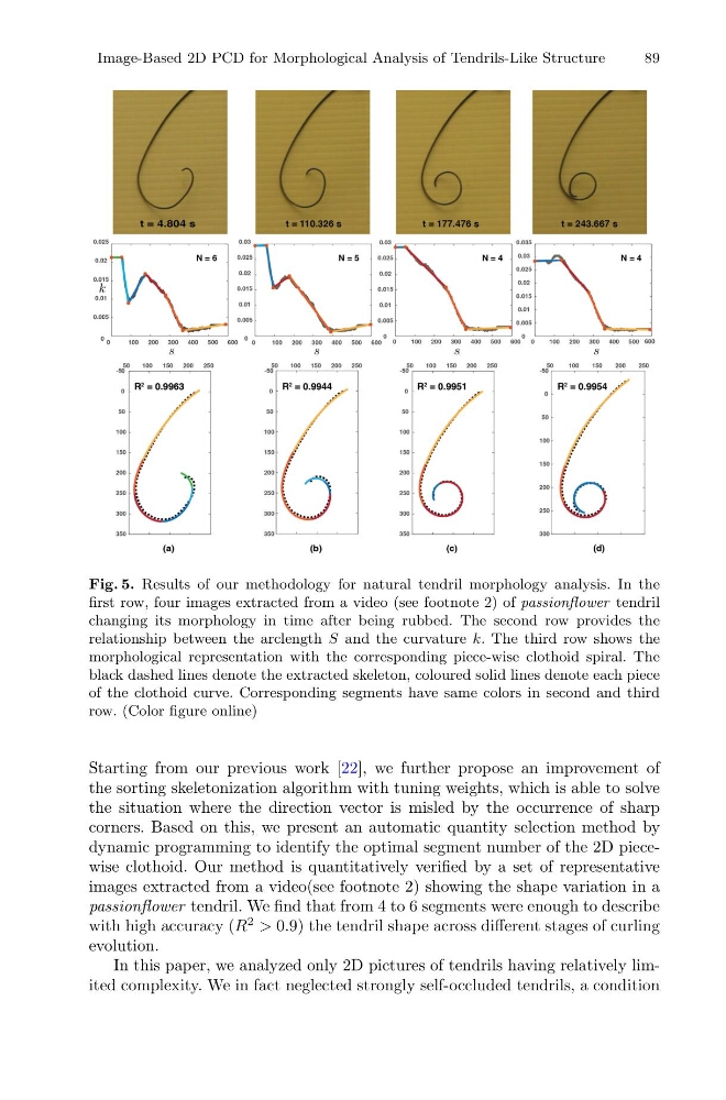
Fig. 5. Results of our methodology for natural tendril morphology analysis. In the
first row, four images extracted from a video (see footnote 2) of passionflower tendril
changing its morphology in time after being rubbed. The second row provides the
relationship between the arclength S and the curvature k. The third row shows the
morphological representation with the corresponding piece-wise clothoid spiral. The
black dashed lines denote the extracted skeleton, coloured solid lines denote each piece
of the clothoid curve. Corresponding segments have same colors in second and third
row. (Color figure online)
Starting from our previous work [22], we further propose an improvement of
the sorting skeletonization algorithm with tuning weights, which is able to solve
the situation where the direction vector is misled by the occurrence of sharp
corners. Based on this, we present an automatic quantity selection method by
dynamic programming to identify the optimal segment number of the 2D piecewise clothoid. Our method is quantitatively verified by a set of representative
images extracted from a video(see footnote 2) showing the shape variation in a
passionflower tendril. We find that from 4 to 6 segments were enough to describe
with high accuracy (R
2 > 0.9) the tendril shape across different stages of curling
evolution.
In this paper, we analyzed only 2D pictures of tendrils having relatively limited complexity. We in fact neglected strongly self-occluded tendrils, a condition
89
Fig. 5. Results of our methodology for natural tendril morphology analysis. In the
first row, four images extracted from a video (see footnote 2) of passionflower tendril
changing its morphology in time after being rubbed. The second row provides the
relationship between the arclength S and the curvature k. The third row shows the
morphological representation with the corresponding piece-wise clothoid spiral. The
black dashed lines denote the extracted skeleton, coloured solid lines denote each piece
of the clothoid curve. Corresponding segments have same colors in second and third
row. (Color figure online)
Starting from our previous work [22], we further propose an improvement of
the sorting skeletonization algorithm with tuning weights, which is able to solve
the situation where the direction vector is misled by the occurrence of sharp
corners. Based on this, we present an automatic quantity selection method by
dynamic programming to identify the optimal segment number of the 2D piecewise clothoid. Our method is quantitatively verified by a set of representative
images extracted from a video(see footnote 2) showing the shape variation in a
passionflower tendril. We find that from 4 to 6 segments were enough to describe
with high accuracy (R
2 > 0.9) the tendril shape across different stages of curling
evolution.
In this paper, we analyzed only 2D pictures of tendrils having relatively limited complexity. We in fact neglected strongly self-occluded tendrils, a condition