

78
C.-L. Hsieh
ing of native biological nanoparticles at ultrahigh speed. Moreover, they illustrated
successful live-cell background removal for single particle dynamics on the cell
surface and inside the cell.
3.4.1 Single Virus Dynamics on Cell Membrane
Using the 3D tracking capability of COBRI (Sect. 3.2) and cell background correction
(Sect. 3.3), highly diffusive motion of a single vaccinia virus particle was revealed
on a live HeLa cell membrane captured at an ultrahigh speed of 100,000 fps. The
virus particles were locally delivered by a micropipette placed close to the cell under
observation. The virus landing process was recorded continuously for a few seconds.
The goal of the experiment was to resolve the early interaction between the virus
particle and the cell plasma membrane receptors immediately after attachment.
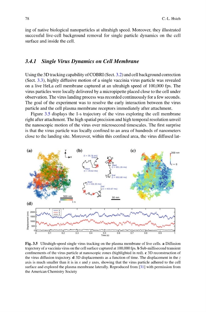
Figure 3.5 displays the 1-s trajectory of the virus exploring the cell membrane
right after attachment. The high spatial precision and high temporal resolution unveil
the nanoscopic motion of the virus over microsecond timescales. The first surprise
is that the virus particle was locally confined to an area of hundreds of nanometers
close to the landing site. Moreover, within this confined area, the virus diffused latFig. 3.5 Ultrahigh-speed single virus tracking on the plasma membrane of live cells. a Diffusion
trajectory of a vaccinia virus on the cell surface captured at 100,000 fps. b Sub-millisecond transient
confinements of the virus particle at nanoscopic zones (highlighted in red). c 3D reconstruction of
the virus diffusion trajectory. d 3D displacements as a function of time. The displacement in the z
axis is much smaller than it is in x and y axes, showing that the virus particle adhered to the cell
surface and explored the plasma membrane laterally. Reproduced from [31] with permission from
the American Chemistry Society
C.-L. Hsieh
ing of native biological nanoparticles at ultrahigh speed. Moreover, they illustrated
successful live-cell background removal for single particle dynamics on the cell
surface and inside the cell.
3.4.1 Single Virus Dynamics on Cell Membrane
Using the 3D tracking capability of COBRI (Sect. 3.2) and cell background correction
(Sect. 3.3), highly diffusive motion of a single vaccinia virus particle was revealed
on a live HeLa cell membrane captured at an ultrahigh speed of 100,000 fps. The
virus particles were locally delivered by a micropipette placed close to the cell under
observation. The virus landing process was recorded continuously for a few seconds.
The goal of the experiment was to resolve the early interaction between the virus
particle and the cell plasma membrane receptors immediately after attachment.
Figure 3.5 displays the 1-s trajectory of the virus exploring the cell membrane
right after attachment. The high spatial precision and high temporal resolution unveil
the nanoscopic motion of the virus over microsecond timescales. The first surprise
is that the virus particle was locally confined to an area of hundreds of nanometers
close to the landing site. Moreover, within this confined area, the virus diffused latFig. 3.5 Ultrahigh-speed single virus tracking on the plasma membrane of live cells. a Diffusion
trajectory of a vaccinia virus on the cell surface captured at 100,000 fps. b Sub-millisecond transient
confinements of the virus particle at nanoscopic zones (highlighted in red). c 3D reconstruction of
the virus diffusion trajectory. d 3D displacements as a function of time. The displacement in the z
axis is much smaller than it is in x and y axes, showing that the virus particle adhered to the cell
surface and explored the plasma membrane laterally. Reproduced from [31] with permission from
the American Chemistry Society