

3 Label-Free, Ultrahigh-Speed, Direct Imaging and Tracking …
77
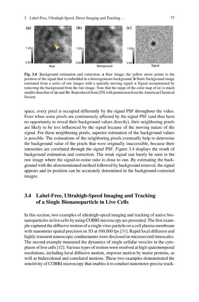
Fig. 3.4 Background estimation and correction. a Raw image; the yellow arrow points to the
position of the signal that is embedded in a heterogeneous background. b Static background image
estimated from a series of raw images with a spatially moving signal. c Signal reconstructed by
removing the background from the raw image. Note that the range of the color map of (c) is much
smaller than that of (a) and (b). Reproduced from [50] with permission from the American Chemical
Society
space, every pixel is occupied differently by the signal PSF throughout the video.
Even when some pixels are continuously affected by the signal PSF (and thus have
no opportunity to reveal their background values directly), their neighboring pixels
are likely to be less influenced by the signal because of the moving nature of the
signal. For these neighboring pixels, superior estimation of the background values
is possible. The estimations of the neighboring pixels eventually help to determine
the background value of the pixels that were originally inaccessible, because their
intensities are correlated through the signal PSF. Figure 3.4 displays the result of
background estimation and correction. The weak signal can barely be seen in the
raw image where the signal-to-noise ratio is close to one. By estimating the background with the aforementioned method followed by background removal, the signal
appears and its position can be accurately determined in the background-corrected
images.
3.4 Label-Free, Ultrahigh-Speed Imaging and Tracking
of a Single Bionanoparticle in Live Cells
In this section, two examples of ultrahigh-speed imaging and tracking of native bionanoparticles in live cells by using COBRI microscopy are presented. The first example captured the diffusive motion of a single virus particle on a cell plasma membrane
with nanometer spatial precision in 3D at 100,000 fps [31]. Rapid local diffusion and
highly transient nanoscopic confinements were disclosed in microsecond timescales.
The second example measured the dynamics of single cellular vesicles in the cytoplasm of live cells [32]. Various types of motion were resolved at high spatiotemporal
resolutions, including local diffusive motion, stepwise motion by motor proteins, as
well as bidirectional and correlated motions. These two examples demonstrated the
sensitivity of COBRI microscopy that enables it to conduct nanometer-precise track-
77
Fig. 3.4 Background estimation and correction. a Raw image; the yellow arrow points to the
position of the signal that is embedded in a heterogeneous background. b Static background image
estimated from a series of raw images with a spatially moving signal. c Signal reconstructed by
removing the background from the raw image. Note that the range of the color map of (c) is much
smaller than that of (a) and (b). Reproduced from [50] with permission from the American Chemical
Society
space, every pixel is occupied differently by the signal PSF throughout the video.
Even when some pixels are continuously affected by the signal PSF (and thus have
no opportunity to reveal their background values directly), their neighboring pixels
are likely to be less influenced by the signal because of the moving nature of the
signal. For these neighboring pixels, superior estimation of the background values
is possible. The estimations of the neighboring pixels eventually help to determine
the background value of the pixels that were originally inaccessible, because their
intensities are correlated through the signal PSF. Figure 3.4 displays the result of
background estimation and correction. The weak signal can barely be seen in the
raw image where the signal-to-noise ratio is close to one. By estimating the background with the aforementioned method followed by background removal, the signal
appears and its position can be accurately determined in the background-corrected
images.
3.4 Label-Free, Ultrahigh-Speed Imaging and Tracking
of a Single Bionanoparticle in Live Cells
In this section, two examples of ultrahigh-speed imaging and tracking of native bionanoparticles in live cells by using COBRI microscopy are presented. The first example captured the diffusive motion of a single virus particle on a cell plasma membrane
with nanometer spatial precision in 3D at 100,000 fps [31]. Rapid local diffusion and
highly transient nanoscopic confinements were disclosed in microsecond timescales.
The second example measured the dynamics of single cellular vesicles in the cytoplasm of live cells [32]. Various types of motion were resolved at high spatiotemporal
resolutions, including local diffusive motion, stepwise motion by motor proteins, as
well as bidirectional and correlated motions. These two examples demonstrated the
sensitivity of COBRI microscopy that enables it to conduct nanometer-precise track-