

2 Interferometric Scattering (iSCAT) Microscopy and Related Techniques
55
Another option for substrate-free membrane studies is to work with giant unilamellar vesicles (GUVs) with diameters in the range of tens of micrometers [179],
which have been pursued as a platform for a minimal cell model [180]. Preliminary results have been reported on tracking of the three-dimensional diffusion of
viral-mimetic particles on the GUV surface, where the particle could be localized to
nanometer precision in all dimensions over an extended range [118], illustrated in
Fig. 2.15b.
2.4.2.3 Imaging Cells and Associated Elements
Three-dimensional monitoring of molecules on the plasma membrane of live cells
to high speed and precision stands as one of the most exciting challenges to unravel
in cell biology [181, 182]. To date, nanoscale transient organization has precluded
satisfactory or compelling investigation by fluorescence methodologies, which fall
short in many respects, especially in capturing three-dimensional landscapes. While
iSCAT is highly suited for addressing this issue, its ultrahigh sensitivity virtue is
accompanied by the vice of a large speckle-like pattern originating from the cellular
membrane and corpus, thus making cellular iSCAT particle tracking a challenge.
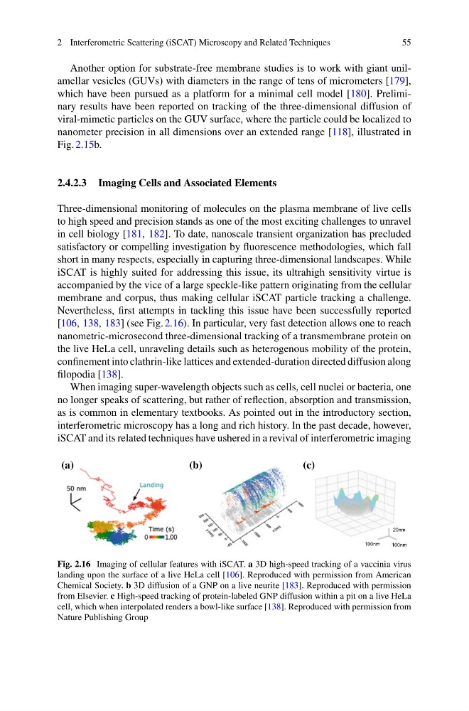
Nevertheless, first attempts in tackling this issue have been successfully reported
[106, 138, 183] (see Fig. 2.16). In particular, very fast detection allows one to reach
nanometric-microsecond three-dimensional tracking of a transmembrane protein on
the live HeLa cell, unraveling details such as heterogenous mobility of the protein,
confinement into clathrin-like lattices and extended-duration directed diffusion along
filopodia [138].
When imaging super-wavelength objects such as cells, cell nuclei or bacteria, one
no longer speaks of scattering, but rather of reflection, absorption and transmission,
as is common in elementary textbooks. As pointed out in the introductory section,
interferometric microscopy has a long and rich history. In the past decade, however,
iSCAT and its related techniques have ushered in a revival of interferometric imaging
100nm
100nm
20nm
(a)
(b)
(c)
Fig. 2.16 Imaging of cellular features with iSCAT. a 3D high-speed tracking of a vaccinia virus
landing upon the surface of a live HeLa cell [106]. Reproduced with permission from American
Chemical Society. b 3D diffusion of a GNP on a live neurite [183]. Reproduced with permission
from Elsevier. c High-speed tracking of protein-labeled GNP diffusion within a pit on a live HeLa
cell, which when interpolated renders a bowl-like surface [138]. Reproduced with permission from
Nature Publishing Group
55
Another option for substrate-free membrane studies is to work with giant unilamellar vesicles (GUVs) with diameters in the range of tens of micrometers [179],
which have been pursued as a platform for a minimal cell model [180]. Preliminary results have been reported on tracking of the three-dimensional diffusion of
viral-mimetic particles on the GUV surface, where the particle could be localized to
nanometer precision in all dimensions over an extended range [118], illustrated in
Fig. 2.15b.
2.4.2.3 Imaging Cells and Associated Elements
Three-dimensional monitoring of molecules on the plasma membrane of live cells
to high speed and precision stands as one of the most exciting challenges to unravel
in cell biology [181, 182]. To date, nanoscale transient organization has precluded
satisfactory or compelling investigation by fluorescence methodologies, which fall
short in many respects, especially in capturing three-dimensional landscapes. While
iSCAT is highly suited for addressing this issue, its ultrahigh sensitivity virtue is
accompanied by the vice of a large speckle-like pattern originating from the cellular
membrane and corpus, thus making cellular iSCAT particle tracking a challenge.
Nevertheless, first attempts in tackling this issue have been successfully reported
[106, 138, 183] (see Fig. 2.16). In particular, very fast detection allows one to reach
nanometric-microsecond three-dimensional tracking of a transmembrane protein on
the live HeLa cell, unraveling details such as heterogenous mobility of the protein,
confinement into clathrin-like lattices and extended-duration directed diffusion along
filopodia [138].
When imaging super-wavelength objects such as cells, cell nuclei or bacteria, one
no longer speaks of scattering, but rather of reflection, absorption and transmission,
as is common in elementary textbooks. As pointed out in the introductory section,
interferometric microscopy has a long and rich history. In the past decade, however,
iSCAT and its related techniques have ushered in a revival of interferometric imaging
100nm
100nm
20nm
(a)
(b)
(c)
Fig. 2.16 Imaging of cellular features with iSCAT. a 3D high-speed tracking of a vaccinia virus
landing upon the surface of a live HeLa cell [106]. Reproduced with permission from American
Chemical Society. b 3D diffusion of a GNP on a live neurite [183]. Reproduced with permission
from Elsevier. c High-speed tracking of protein-labeled GNP diffusion within a pit on a live HeLa
cell, which when interpolated renders a bowl-like surface [138]. Reproduced with permission from
Nature Publishing Group