

52
R. W. Taylor and V. Sandoghdar
(a)
(b)
(c)
Time (s)
Distance (nm)
0.0
1.0
2.0
3.0
4.0
74
148
222
296
370
0
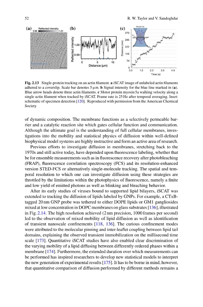
Fig. 2.13 Single-protein tracking on an actin filament. a iSCAT image of unlabeled actin filaments
adhered to a coverslip. Scale bar denotes 5 µm. b Signal intensity for the blue line marked in (a).
Blue arrow heads denote three actin filaments. c Motor protein myosin 5a walking velocity along a
single actin filament when tracked by iSCAT. Frame rate is 25 Hz after temporal averaging. Inset:
schematic of specimen detection [120]. Reproduced with permission from the American Chemical
Society
of dynamic composition. The membrane functions as a selectively permeable barrier and a catalytic reaction site which gates cellular function and communication.
Although the ultimate goal is the understanding of full cellular membranes, investigations into the mobility and statistical physics of diffusion within well-defined
biophysical model systems are highly instructive and form an active area of research.
Previous efforts to investigate diffusion in membranes, stretching back to the
1970s and still active today, have depended upon fluorescence labeling, whether that
be for ensemble measurements such as in fluorescence recovery after photobleaching
(FRAP), fluorescence correlation spectroscopy (FCS) and its resolution-enhanced
version STED-FCS or alternatively single-molecule tracking. The spatial and temporal resolution to which one can investigate diffusion using these strategies are
throttled by the limitations within the photophysics of fluorescence, namely a finite
and low yield of emitted photons as well as blinking and bleaching behavior.
After its early studies of viruses bound to supported lipid bilayers, iSCAT was
extended to tracking the diffusion of lipids labeled by GNPs. For example, a CTxBtagged 20 nm GNP probe was tethered to either DOPE lipids or GM1 gangliosides
mixed at low concentration in DOPC membranes on glass substrates [136], illustrated
in Fig. 2.14. The high resolution achieved (2 nm precision, 1000 frames per second)
led to the observation of mixed mobility of lipid diffusion as well as identification
of transient nanoscale confinements [118, 136]. The curious confinement modes
were attributed to the molecular pinning and inter-leaflet coupling between lipid tail
domains, explaining the observed transient immobilization on the millisecond time
scale [173]. Quantitative iSCAT studies have also enabled clear discrimination of
the varying mobility of a lipid diffusing between differently ordered phases within a
membrane [174]. Furthermore, the extended duration over which measurements can
be performed has inspired researchers to develop new statistical models to interpret
the new generation of experimental results [175]. It has to be borne in mind, however,
that quantitative comparison of diffusion performed by different methods remains a
R. W. Taylor and V. Sandoghdar
(a)
(b)
(c)
Time (s)
Distance (nm)
0.0
1.0
2.0
3.0
4.0
74
148
222
296
370
0
Fig. 2.13 Single-protein tracking on an actin filament. a iSCAT image of unlabeled actin filaments
adhered to a coverslip. Scale bar denotes 5 µm. b Signal intensity for the blue line marked in (a).
Blue arrow heads denote three actin filaments. c Motor protein myosin 5a walking velocity along a
single actin filament when tracked by iSCAT. Frame rate is 25 Hz after temporal averaging. Inset:
schematic of specimen detection [120]. Reproduced with permission from the American Chemical
Society
of dynamic composition. The membrane functions as a selectively permeable barrier and a catalytic reaction site which gates cellular function and communication.
Although the ultimate goal is the understanding of full cellular membranes, investigations into the mobility and statistical physics of diffusion within well-defined
biophysical model systems are highly instructive and form an active area of research.
Previous efforts to investigate diffusion in membranes, stretching back to the
1970s and still active today, have depended upon fluorescence labeling, whether that
be for ensemble measurements such as in fluorescence recovery after photobleaching
(FRAP), fluorescence correlation spectroscopy (FCS) and its resolution-enhanced
version STED-FCS or alternatively single-molecule tracking. The spatial and temporal resolution to which one can investigate diffusion using these strategies are
throttled by the limitations within the photophysics of fluorescence, namely a finite
and low yield of emitted photons as well as blinking and bleaching behavior.
After its early studies of viruses bound to supported lipid bilayers, iSCAT was
extended to tracking the diffusion of lipids labeled by GNPs. For example, a CTxBtagged 20 nm GNP probe was tethered to either DOPE lipids or GM1 gangliosides
mixed at low concentration in DOPC membranes on glass substrates [136], illustrated
in Fig. 2.14. The high resolution achieved (2 nm precision, 1000 frames per second)
led to the observation of mixed mobility of lipid diffusion as well as identification
of transient nanoscale confinements [118, 136]. The curious confinement modes
were attributed to the molecular pinning and inter-leaflet coupling between lipid tail
domains, explaining the observed transient immobilization on the millisecond time
scale [173]. Quantitative iSCAT studies have also enabled clear discrimination of
the varying mobility of a lipid diffusing between differently ordered phases within a
membrane [174]. Furthermore, the extended duration over which measurements can
be performed has inspired researchers to develop new statistical models to interpret
the new generation of experimental results [175]. It has to be borne in mind, however,
that quantitative comparison of diffusion performed by different methods remains a