

2 Interferometric Scattering (iSCAT) Microscopy and Related Techniques
27
B
r
i
g
h
t
-
f
i
e
l
d
m
i
c
r
o
s
c
o
p
y
16
th
Century
Today
i n t e r f e r o m
e t r i c
s c a t t e r i n g
m
i c r o s c o p y
( i S C A T )
L a s e r
i n v e n t e d
P h o t o g r a p h i c
i m
a g i n g
A n a l o g u e
v i d e o
H
o l o g r a p h y
P h a s e
c o n t r a s t
m
i c r o s c o p y
C o n f o c a l
m
i c r o s c o p y D
I C
m
i c r o s c o p y
V i d e o - e n h a n c e d
m
i c r o s c o p y D
i g i t a l v i d e o
D
a
r
k
-
f
i
e
l
d
m
i
c
r
o
s
c
o
p
y
S u p e r - r e s o l u t i o n
f l u o r e s c e n c e
m
i c r o s c o p y
20
th
Century
19
th
Century
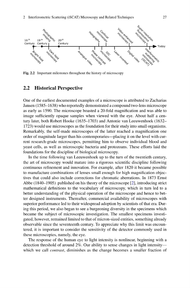
Fig. 2.2 Important milestones throughout the history of microscopy
2.2 Historical Perspective
One of the earliest documented examples of a microscope is attributed to Zacharias
Jansen (1585–1638) who reportedly demonstrated a compound two-lens microscope
as early as 1590. The microscope boasted a 20-fold magnification and was able to
image sufficiently opaque samples when viewed with the eye. About half a century later, both Robert Hooke (1635–1703) and Antonie van Leeuwenhoek (1632–
1723) would use microscopes as the foundation for their study into small organisms.
Remarkably, the self-made microscopes of the latter reached a magnification one
order of magnitude larger than his contemporaries—placing it on the level with current research-grade microscopes, permitting him to observe individual blood and
yeast cells, as well as microscopic bacteria and protozoans. These efforts laid the
foundations for the discipline of biological microscopy.
In the time following van Leeuwenhoek up to the turn of the twentieth century,
the art of microscopy would mature into a rigorous scientific discipline following
continuous refinement and innovation. For example, after 1820 it became possible
to manufacture combinations of lenses small enough for high magnification objectives that could also include corrections for chromatic aberrations. In 1873 Ernst
Abbe (1840–1905) published on his theory of the microscope [2], introducing strict
mathematical definitions to the vocabulary of microscopy, which in turn led to a
better understanding of the physical operation of the microscope and hence to better designed instruments. Thereafter, commercial availability of microscopes with
superior performance led to their widespread adoption by scientists of that era. During this period, we also began to see a burgeoning diversity in the specimens which
became the subject of microscopic investigation. The smallest specimens investigated, however, remained limited to that of micron-sized entities, something already
observable since the seventeenth century. To appreciate why this limit was encountered, it is important to consider the sensitivity of the detector commonly used in
these microscopies, namely, the eye.
The response of the human eye to light intensity is nonlinear, beginning with a
detection threshold of around 2%. Our ability to sense changes in light intensity—
which we call contrast, diminishes as the change becomes a smaller fraction of
27
B
r
i
g
h
t
-
f
i
e
l
d
m
i
c
r
o
s
c
o
p
y
16
th
Century
Today
i n t e r f e r o m
e t r i c
s c a t t e r i n g
m
i c r o s c o p y
( i S C A T )
L a s e r
i n v e n t e d
P h o t o g r a p h i c
i m
a g i n g
A n a l o g u e
v i d e o
H
o l o g r a p h y
P h a s e
c o n t r a s t
m
i c r o s c o p y
C o n f o c a l
m
i c r o s c o p y D
I C
m
i c r o s c o p y
V i d e o - e n h a n c e d
m
i c r o s c o p y D
i g i t a l v i d e o
D
a
r
k
-
f
i
e
l
d
m
i
c
r
o
s
c
o
p
y
S u p e r - r e s o l u t i o n
f l u o r e s c e n c e
m
i c r o s c o p y
20
th
Century
19
th
Century
Fig. 2.2 Important milestones throughout the history of microscopy
2.2 Historical Perspective
One of the earliest documented examples of a microscope is attributed to Zacharias
Jansen (1585–1638) who reportedly demonstrated a compound two-lens microscope
as early as 1590. The microscope boasted a 20-fold magnification and was able to
image sufficiently opaque samples when viewed with the eye. About half a century later, both Robert Hooke (1635–1703) and Antonie van Leeuwenhoek (1632–
1723) would use microscopes as the foundation for their study into small organisms.
Remarkably, the self-made microscopes of the latter reached a magnification one
order of magnitude larger than his contemporaries—placing it on the level with current research-grade microscopes, permitting him to observe individual blood and
yeast cells, as well as microscopic bacteria and protozoans. These efforts laid the
foundations for the discipline of biological microscopy.
In the time following van Leeuwenhoek up to the turn of the twentieth century,
the art of microscopy would mature into a rigorous scientific discipline following
continuous refinement and innovation. For example, after 1820 it became possible
to manufacture combinations of lenses small enough for high magnification objectives that could also include corrections for chromatic aberrations. In 1873 Ernst
Abbe (1840–1905) published on his theory of the microscope [2], introducing strict
mathematical definitions to the vocabulary of microscopy, which in turn led to a
better understanding of the physical operation of the microscope and hence to better designed instruments. Thereafter, commercial availability of microscopes with
superior performance led to their widespread adoption by scientists of that era. During this period, we also began to see a burgeoning diversity in the specimens which
became the subject of microscopic investigation. The smallest specimens investigated, however, remained limited to that of micron-sized entities, something already
observable since the seventeenth century. To appreciate why this limit was encountered, it is important to consider the sensitivity of the detector commonly used in
these microscopies, namely, the eye.
The response of the human eye to light intensity is nonlinear, beginning with a
detection threshold of around 2%. Our ability to sense changes in light intensity—
which we call contrast, diminishes as the change becomes a smaller fraction of